Monday, July 31, 2017

Parasite Case of the Week 454

This week's case was generously donated by Florida Fan. The patient is an otherwise well woman in her 30's who passed the following object in her stool:


The following object was also observed in her stool. It measured approximately 50 micrometers in length. Identification?

Sunday, July 30, 2017

Answer to Case 454

Answer: Tapeworm, Diphyllobothrium species

This case shows the classic morphology of an adult tapeworm, consisting of a flat ribbon-like body composed of multiple repeating units called proglottids. This particular tapeworm can be identified by the morphology of its proglottids and eggs. The proglottids are broader/wider than they are long; hence the name the "broad fish tapeworm". Within the center of each proglottid is a central uterine structure which is often visible macroscopically. In this case, it can be seen as a central 'hump' in each proglottid:
The eggs are also characteristic for this genus and allow for differentiation between Diphyllobothrium and Taenia species. Note the thin wall, oval shape, intermediate size (55-75 micrometers long) of the egg and the operculum (lid-like opening) at one end. In many cases, a knob-like structure can be seen at the other end (abopercular knob), but it is not clearly visible in this case.
The most reliable means for identifying the infecting Diphyllobothrium species is through molecular testing. While D. latum is the most common cause of human infection worldwide, other species that can infect humans include D. pacificum, D. yonagoensis, D. dendriticum, D. dalliae, D. cordatum, D. ursi, and D. lanceolatum.

Saturday, July 22, 2017

Case of the Week 453

This week's case was kindly donated by one of my former Clinical Microbiology fellows, Dr. Adam Caulfield. The specimen (shown below) was extracted from the leg of a 70 year old man who had recently returned from a trip to Costa Rica. It measures approximately 1.5 cm in length. Identification?

Friday, July 21, 2017

Answer to Case 453

Answer: Bot fly, Dermatobia hominis

As mentioned by Florida Fan, the body has spiky projections and general body habitus consistent with myiasis. Of note, the cuticular spines are lacking on the terminal 3 body segments which, along with the travel history, are consistent with Dermatobia hominis. Ideally we would also like to look at the posterior spiracles to confirm the identification, but photos weren't available from this case.

Tuesday, July 11, 2017

Case of the Week 452

This week's case was generously donated by Dr. Richard Bradbury. The following biphasic population of organisms was seen after corneal scrapings were inoculated onto a non-nutrient agar overlain with Escherichia coli and incubated for several days.

These objects measure approximately 25 micrometers in diameter.


These objects measured up to 45 micrometers in greatest dimension:


Identification?

Monday, July 10, 2017

Answer to Case 452

Answer: 
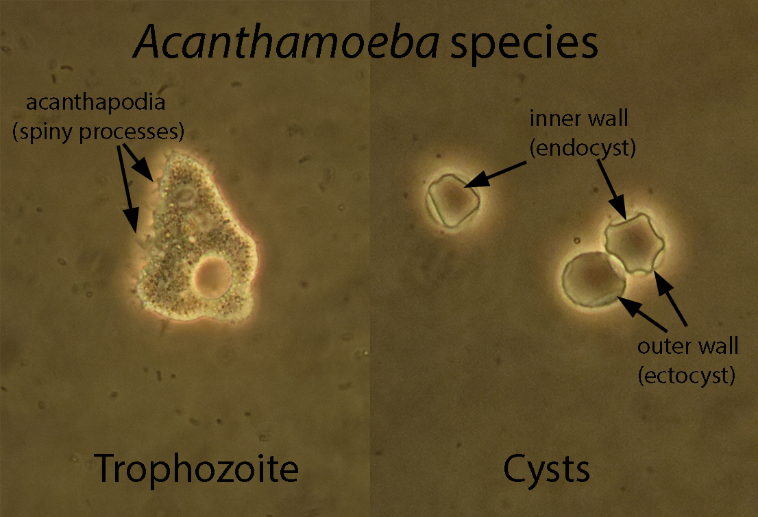
Acanthamoeba species cysts and trophozoites. The diagnosis can be made by the characteristic morphology in conjunction with the clinical history. As mentioned by Florida Fan, the trophozoites have thorny or spiny projections called acanthopodia (from the Greek akantha meaning thorn), whereas the trophozoites of other amebae have rounded ends on their pseudopodia/projections.

The source (cornea) is also characteristic for this organism. Richard Garcia-Kennedy mentioned that not changing the contact lens case regularly is a common risk factor. Other important risk factors are cleaning contact lenses (or the case) with tap water and swimming while wearing lenses. I think I've scared all of my residents and Clinical Microbiology fellows out of these bad habits!

Like Rune, we have switched over to PCR for detection of all free-living amebae from clinical specimens. However, we used to use tap water agar (overlain with a bacterium like E. coli as a food source), and we would also occasionally supply our "cornealogists" with culture plates that they could directly inoculate. However we mostly had our clinicians send us specimens in MEM (minimum essential medium) which also worked quite well in supporting growth and viability until we could inoculate the specimen onto culture. When positive, corneal specimens and contact lenses usually grew Acanthamoeba within 24 hours (and in one memorable case, within 8 hours), but the PCR provides an even faster result and has proven to be just as sensitive as culture. It also allows us to detect Balamuthia mandrillaris (which won't grow on non-nutrient agar) and Naegleria fowleri (where time is of the essence for detection).

Thank you all for the comments on this case, and thanks again to Richard for donating it!

Monday, July 3, 2017

Case of the week 451

Happy July 4th to my American readers! Here is a nice red, white and blue case for you. The following objects were seen in a stool specimen stained with a modified acid fast stain. They measure approximately 35 micrometers in greatest dimension. Identification?


Sunday, July 2, 2017

Answer to Case 451

Answer: Cystoisospora belli (formerly Isospora belli) oocysts

As with Cyclospora cayetanensis and Cryptosporidium oocysts, the oocysts of C. belli stain red/pink using modified acid fast and modified safranin stains (below, right), with unstained "ghost" cells (below, left) also commonly seen. The oocysts are immature when shed in stool and contain only a single sporoblast. Oocysts will then mature in the soil, during which time the sporoblast divides into 2 sporoblasts  and develop cyst walls, becoming sporocysts. Each sporocyst will contain 4 sporozoites - the form that invades the intestinal epithelial cells.