Tuesday, February 28, 2017
Case of the Week 436
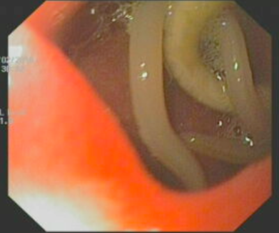
This week's case was generously donated by Dr. Sabarinathan from Madras Medical College in Chennai, India. The patient is a young man with a 2-week history of epigastric pain and vomiting. Physical examination was normal, and a complete blood count showed only a mild anemia (hemoglobin of 9.8 g/dL). An ultrasound of the abdomen was normal and so an upper gastrointestinal endoscopy was performed. This is what was seen:
Identification?
Monday, February 27, 2017
Answer to Case 436
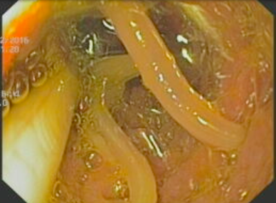
Answer: Ascaris lumbricoides
Although we don't have the worms to examine in this case, the presence of multiple large tan-white worms with a smooth outer cuticle within the lumen of the intestine is strongly consistent with A. lumbricoides. The female A. lumbricoides can get quite large, reaching lengths of 35 cm!
One reader also raised the possibility of anisakiasis given that these worms were found on an upper endoscopy. However, the large size would exclude anisakid larvae given that they only reach a few centimeters in length. Also, the presence of multiple worms would be unlikely for anisakiasis, since usually only 1 larva is seen in these cases. Finally, anisakids try to recapitulate their lifecycle in their marine mammal host and embed into the gastric or intestinal mucosa. This type of behavior was not seen in this case.
Although we don't have the worms to examine in this case, the presence of multiple large tan-white worms with a smooth outer cuticle within the lumen of the intestine is strongly consistent with A. lumbricoides. The female A. lumbricoides can get quite large, reaching lengths of 35 cm!


One reader also raised the possibility of anisakiasis given that these worms were found on an upper endoscopy. However, the large size would exclude anisakid larvae given that they only reach a few centimeters in length. Also, the presence of multiple worms would be unlikely for anisakiasis, since usually only 1 larva is seen in these cases. Finally, anisakids try to recapitulate their lifecycle in their marine mammal host and embed into the gastric or intestinal mucosa. This type of behavior was not seen in this case.
Sunday, February 19, 2017
Case of the Week 435
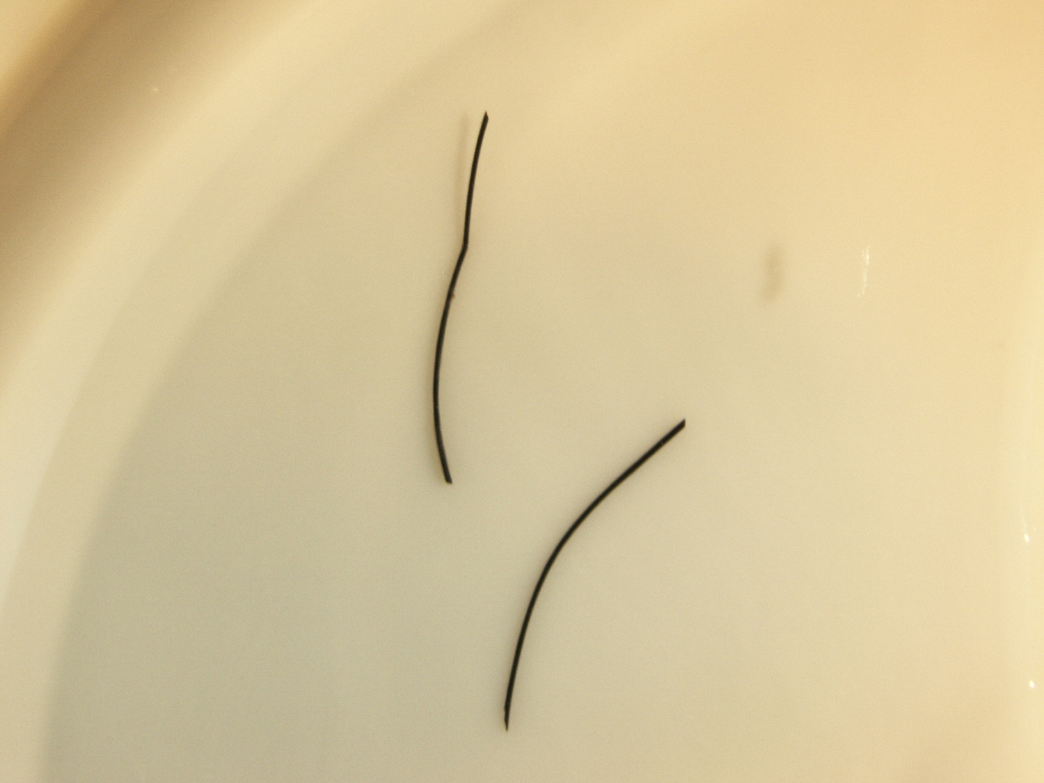
This week's challenging case was donated by Dr. Emily Hall. The patient is a previously-healthy toddler who began acting unusually fussy and refused to ambulate. Examination reviewed a thin black object under the skin of her right foot. Extraction was unsuccessful and the child was playing and ambulating at the end of the visit and was therefore sent home. The child was brought back the following day because the object had moved approximately 3 cm within a 24 hour period in the semilunar pattern shown in the image below:
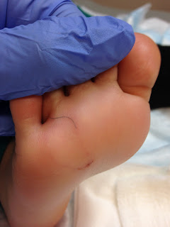 At this point, the object was removed and sent to the parasitology lab. Rather than showing you the images of the object that was extracted, I thought I would ask for your diagnosis based on the clinical image alone. I will show you the images and provide the diagnosis next week!
The image is shown with permission from the mother.

The image is shown with permission from the mother.
Saturday, February 18, 2017
Answer to Case 435
Wow, this case generated a lot of interest, with requests for the answer when I didn't post yesterday. My apologies for leaving you all hanging!
This is a case of a non-parasitic fiber that had been transcutaneously implanted and migrated under the skin due to physical pressure (i.e. walking). Although the presentation was suggestive of a cutaneous parasitic infection such as cutaneous larva migrans (CLM), the length of the object and dark color were not consistent with any human or zoonotic parasite. Several readers pointed out specifically that the larvae that cause CLM are microscopic and therefore would not be visible to the naked eye. Also, the path formed by a migrating worm would be more serpiginous rather than the semi-lunar pattern observed here. Other subcutaneous or intra-epidermal/dermal worms such as Loa loa, zoonotic microfilariae, and Dracunculus medinensis were also suggested but rightly discarded by readers because of the size and color of this object. There is no parasite, to my knowledge, that would have this appearance and be present in this location in human skin.
One intriguing suggestion was that this was cutaneous pili migrans (CPM) - an excellent thought. CPM is a phenomenon in which a hair grows within the skin rather than up and out of the skin, and is seen as long dark object just below the skin's surface. This is most common on hair-bearing regions of the body and would be unusual on the sole of the foot. However, removal of the object would be required to rule out CPM. Therefore, this is exactly what we did. Here are photographs of the object that we removed (cut in half):

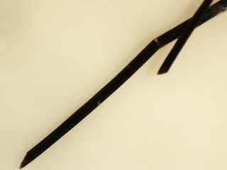 As you can see, it looks to be a synthetic fiber and not any type of worm. It was extremely resilient, and while bendable, did not easily break. Just for fun (and because I knew you would all want to know), we analyzed the fiber using infrared spectrum analysis, which showed it to be most consistent with azlon, a synthetic fiber commonly used to make clothing and household objects.
Thanks again to Emily Hall for donating this fascinating case!
This is a case of a non-parasitic fiber that had been transcutaneously implanted and migrated under the skin due to physical pressure (i.e. walking). Although the presentation was suggestive of a cutaneous parasitic infection such as cutaneous larva migrans (CLM), the length of the object and dark color were not consistent with any human or zoonotic parasite. Several readers pointed out specifically that the larvae that cause CLM are microscopic and therefore would not be visible to the naked eye. Also, the path formed by a migrating worm would be more serpiginous rather than the semi-lunar pattern observed here. Other subcutaneous or intra-epidermal/dermal worms such as Loa loa, zoonotic microfilariae, and Dracunculus medinensis were also suggested but rightly discarded by readers because of the size and color of this object. There is no parasite, to my knowledge, that would have this appearance and be present in this location in human skin.
One intriguing suggestion was that this was cutaneous pili migrans (CPM) - an excellent thought. CPM is a phenomenon in which a hair grows within the skin rather than up and out of the skin, and is seen as long dark object just below the skin's surface. This is most common on hair-bearing regions of the body and would be unusual on the sole of the foot. However, removal of the object would be required to rule out CPM. Therefore, this is exactly what we did. Here are photographs of the object that we removed (cut in half):

Thanks again to Emily Hall for donating this fascinating case!
Saturday, February 11, 2017
Case of the Week 434
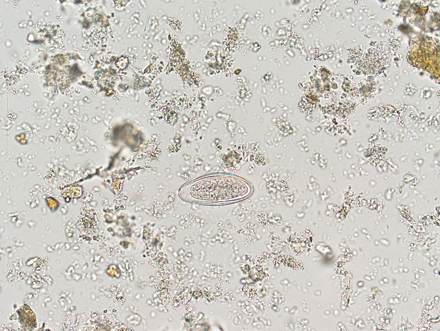
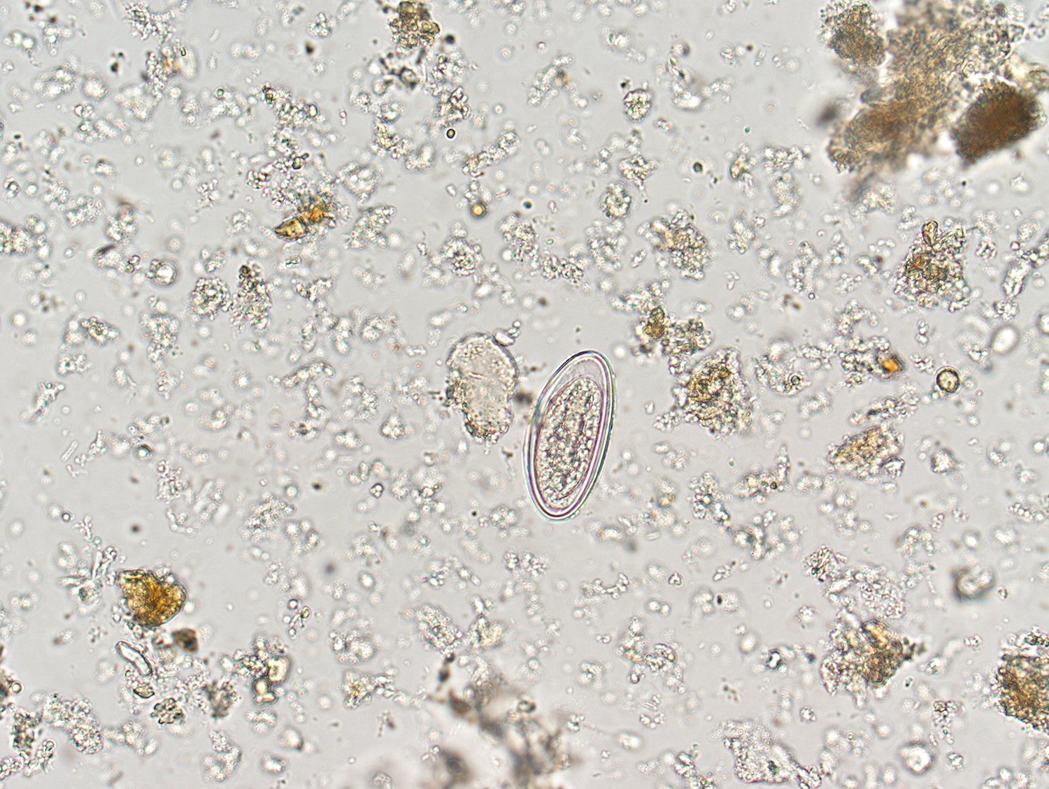
This week's case is another simple identification. The following structures were seen in stool of a young child.


What are these structures?

What are these structures?
Friday, February 10, 2017
Answer to Case 434
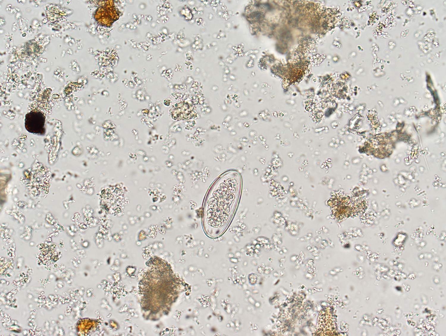
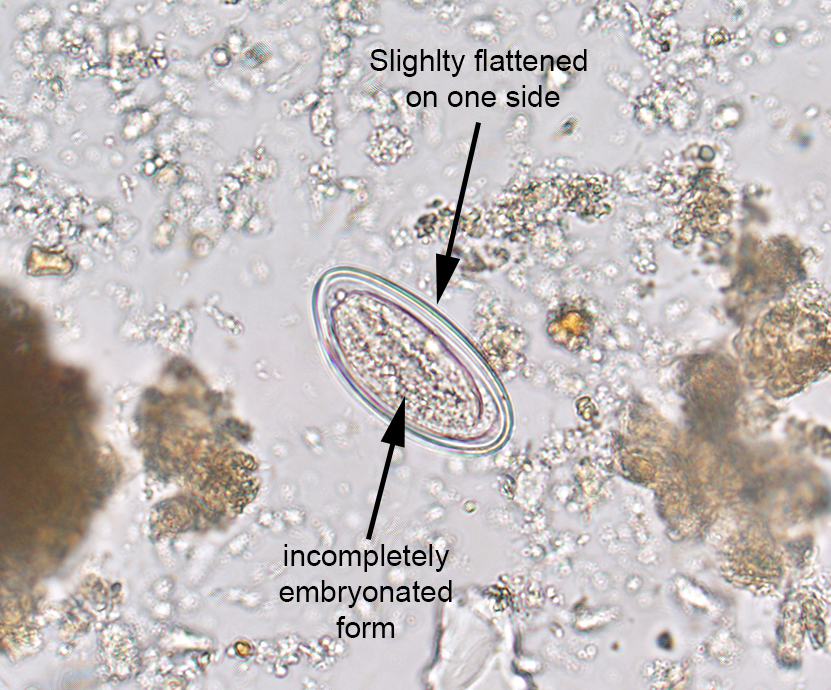
Answer: Enterobius vermicularis (pinworm) eggs
Note the characteristic features including the thin colorless shell and partially-oval shape that is flattened on one side. The larvae are not fully developed within these eggs, but generally are developed (and infective) within 4-6 hours of the eggs being laid.
 This was an interesting case in that the eggs were seen in stool rather on a peri-anal adhesive tape preparation. The latter is the preferred method for detecting pinworm eggs since the adult female lays her eggs on the perianal skin folds. However, eggs may attach to the stool as it passes through the anus, and are therefore occasionally (but not reliably!) seen using the ova and parasite exam.
Note the characteristic features including the thin colorless shell and partially-oval shape that is flattened on one side. The larvae are not fully developed within these eggs, but generally are developed (and infective) within 4-6 hours of the eggs being laid.
Sunday, February 5, 2017
Case of the Week 433
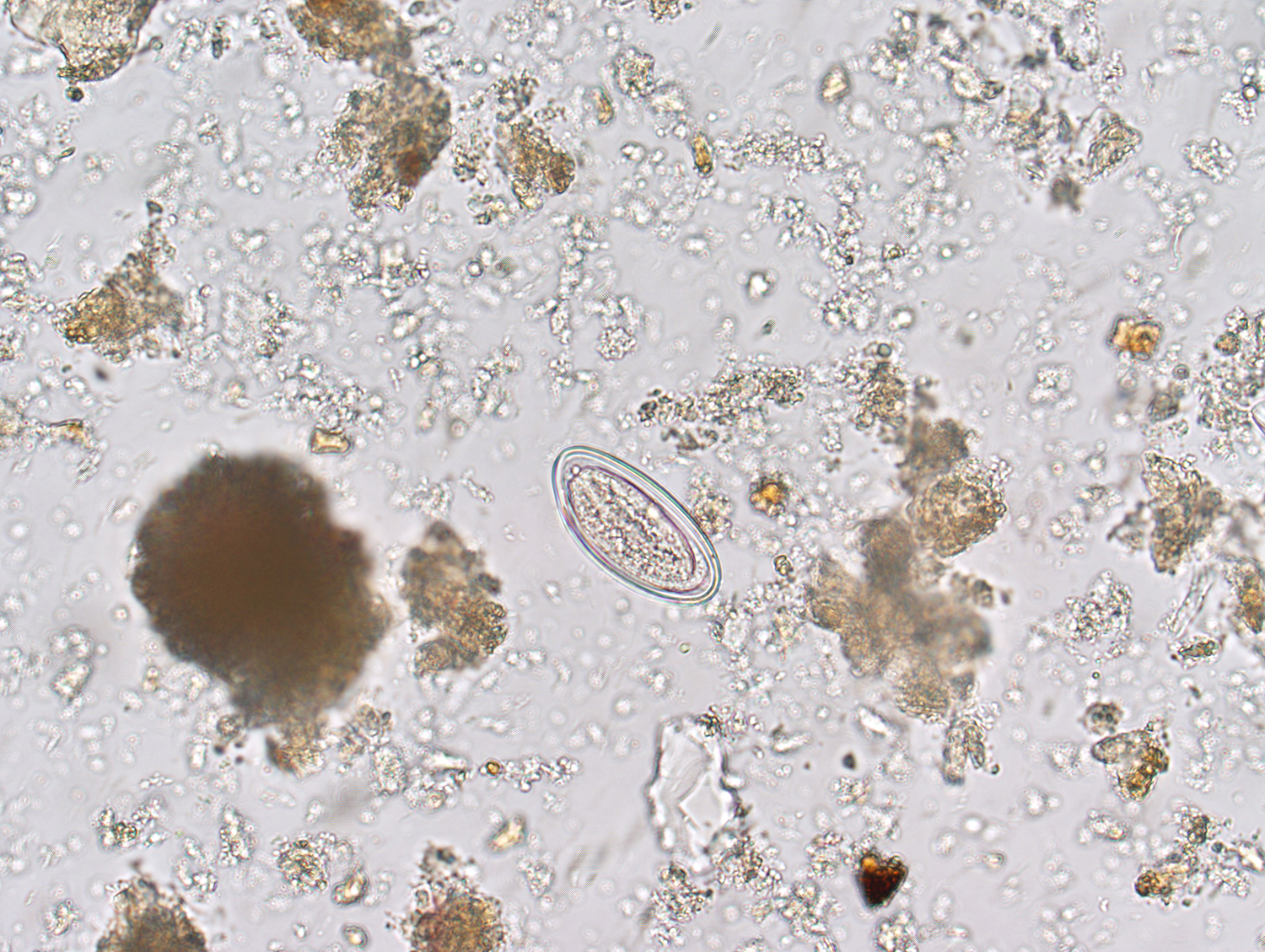
This week's images were generously donated by Florida Fan. The following structures were seen in a concentrated stool specimen and measured approximately 75 micrometers in greatest dimension (2.5 micrometers per line on the scale bar). Identification?


Saturday, February 4, 2017
Answer to Case 433
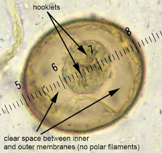
Answer: Hymenolepis diminuta, the rat tapeworm. Humans become infected when accidentally ingesting infected arthropods.
H. diminuta eggs can be differentiated from the similar-appearing eggs of Hymenolepis nana by their larger size (70-80 micrometers in greatest dimension) and lack of polar filaments. In contrast, H. nana eggs are 30-50 micrometers in greatest dimension and have polar filaments radiating out from the inner shell. Both eggs have an inner shell surrounding the 6-hooked onchosphere and an outer shell.
Here are some of the defining features of H. diminuta eggs:

Subscribe to:
Posts (Atom)


