Congratulations to Martin, Ali, Florida Fan, Mark, Atiya, Sara, and Alexandra who got this correct!
The videos show the beautiful 'spiraling' motility of this organism, similar but distinct from the 'falling leaf' motility of Giardia and the 'jerky' motility of Pentatrichomonas hominis. In the lab, of course, we would also have the final fixed morphology to aid in our diagnosis and confirm our impression from the direct preparation.
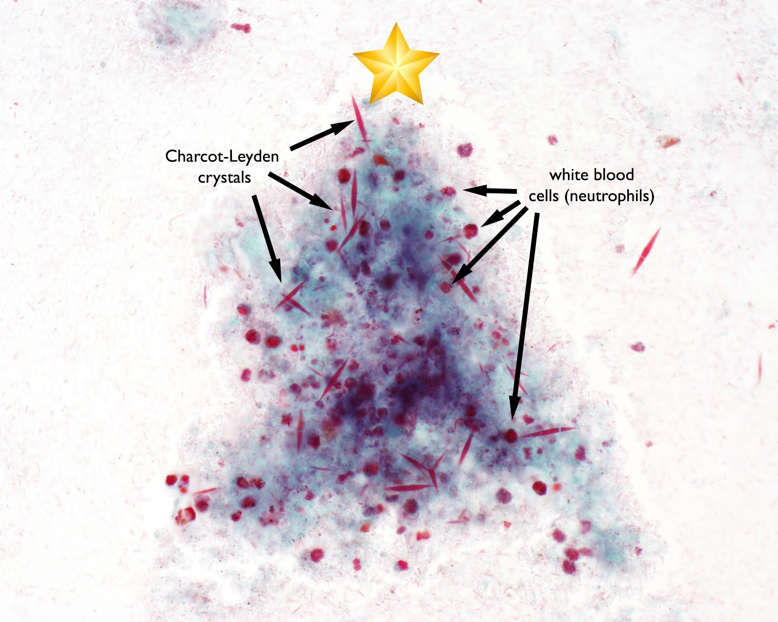
Happy Holidays to all of my readers! This week's case is in the form of a Christmas tree. Can anyone tell what the tree is made out of? (hint: this is from a Trichrome-stained stool specimen)
Answer: Charcot-Leyden crystals, white blood cells and red blood cells
As Florida Fan mentioned, the C-L crystals are a guise for snow crystals, and the red cells may perhaps represent red delicious apples before man-made ornaments were innovated. I also envision the C-L crystals representing pine needles, perhaps?
I was very pleased to stumble upon this 'tree' when looking for some crystals to photograph. Other than the star, nothing else has been digitally added to this image.
Answer: Blastocystis sp. AND pollen.
Wow, you all impressed me by noting the less obvious Blastocystis in image 1, in addition to the pollen in all 3 images. Also, no one mistakenly thought that the pollen was a helminth egg (e.g. Taenia sp.) - a common pitfall. Excellent job!
While the Blastocystis cyst-like forms are the only parasites presents (image 1), I do agree that the pollen is the most striking finding. And very festive as Florida Fan and Idzi mentioned! I will defer the identification of the pollen to the others as this is outside of my area of expertise, but appreciate the time that some readers took to identifying the specific variety shown here.
Answer: Hard tick (Ixodidae) nymph emerging from the larval exuvia. The inverted "U-shaped" anal groove on the ventral surface (photo below) allows us to identify this as an Ixodes species.
The gender is not possible to discern in nymphs, but as mentioned by Anon, it can be determined in adults by examining the scutum and basis capituli. The scutum (dorsal shield) of the female only covers a portion of the dorsal surface, compared to the male in which it covers nearly the entire dorsal surface. In the nymphal stage, both males and females have a similar-appearing scutum. Also, only females have porose areas whereas males do not. The porose areas are located on either side of the basis capituli and produce antioxidants which are combined with waxy secretions from the Gene's organ. This substance is applied to eggs right after they are laid and serves as a protective coating. The porose areas also lubricate the Gene's organ, allowing it to expand and retract more easily.
Thanks again to Dr. Graham Hickling for donating this fascinating video. Thank you also to Dr. Robyn Nadolny for the additional information about the function of porose areas and Gene's organ. If you'd like to see the Gene's organ in action (and tick eggs being laid), check out this other amazing video by Graham.
Welcome to the first case of the month featuring a case from Idzi Potters at the Institute of Tropical Medicine Antwerp. This case is really spectacular and something we don't see very often.
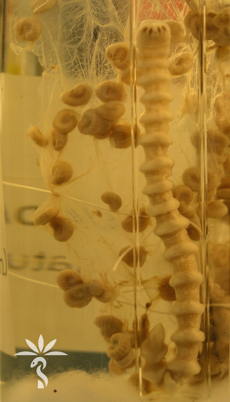
The following parasites were discovered in a man's peritoneal tissue during an inguinoscrotal hernia repair. The resided in Benin, Africa. Here is the resected section of peritoneum along with the attached parasites (CLICK ON IMAGE TO ENLARGE):
Answer: Pentostomiasis; most consistent with Armillifer species. William Sears also suggested that Raillietiella sp. is also in the differential, which Idzi confirms. Both are found in African countries and associated with consumption of raw snake meat. Identification is accomplished morphologically by counting the annuli. You can also use molecular studies, although these are not widely available.
Idzi also provided me with the following beautiful (and very creepy) photos from his Institute's specimen archives which show an adult and larval Armillifer armillatus:
Idzi and his group published this case, so you can read more about it HERE. Fascinating case!
This week's case was donated from Dr. Kamran Kadkhoda. This worm was submitted to the laboratory in saline. It had been seen on the surface of stool from a 3 year old girl.
The following objects were seen within the worm and in the saline submitted with the specimen. They measure approximately 60 micrometers in length.
Answer: Enterobius vermicularis (or E. gregorii); a.k.a. pinworm.
Several of you noted the classic features of the female pinworm shown in this case: the prominent anterior cervical alae, classic eggs (in and outside of the worm), and the slender "pin-like" tail that gives this worm its common name. Males also prominent cervical alae but lack the pointy tail; instead they have a blunt, often curved, posterior end with a single spicule.
As mentioned by Florida Fan, infected patients typically experience nocturnal anal pruritus, and the worm may be observed crawling on the surface of the stool. Ali Mokbel also noted that each work lays approximately 10,000 eggs each day. Importantly, these eggs are fully infectious within 4-6 hours of being laid, and this is one of the most important reasons why this worm is common in the United States and other resource-rich temperate climates. The eggs of most other intestinal nematodes require an incubation period in the soil before becoming infectious, and therefore infection can be prevented with proper sanitation measures, including waste treatment.
Thank you again to Dr. Kadkhoda for donating this classic case!
This is a special post to wish a very happy Thanksgiving to all of my American readers. Can you all see the turkey head in the following blood smear? (you may need to use your imagination a bit).
The image is courtesy of my awesome lab education specialist Emily Fernholz. Can you tell what Plasmodium species is shown here?
The answer to Case of the Week 469 will be posted tomorrow.
The below was seen on a stool agar culture after incubation at room temperature for several days. The patient is a 62-year-old woman from the Philippines. The images are by my awesome lead tech, Heather Rose, while the video is by Emily Fernholz, Education Specialist extraordinare.
The following were seen in the concentrated wet preparation of the stool specimen :
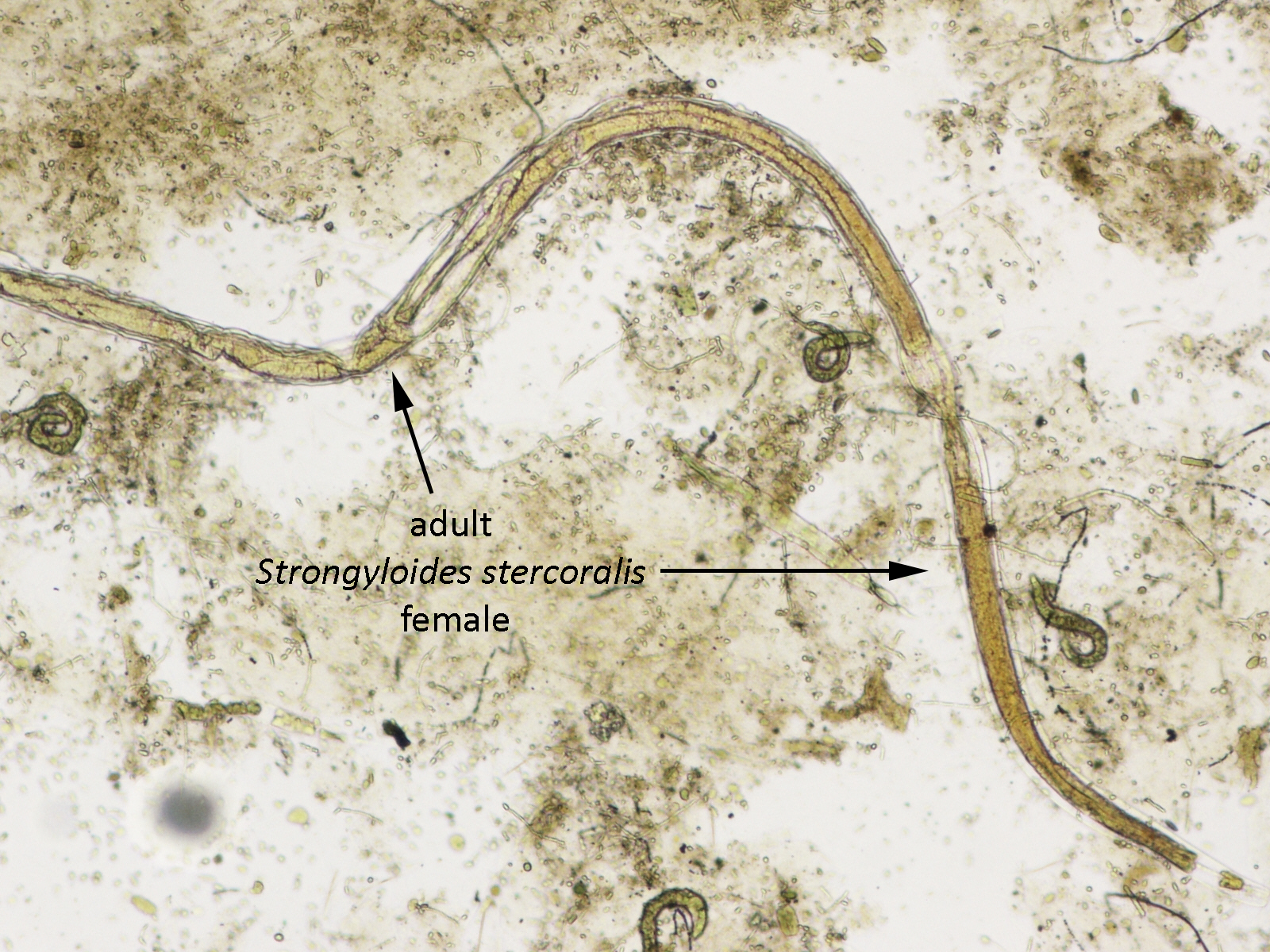
Answer: Strongyloides stercoralis. Note the characteristic morphology and the impressive larval load in the stool agar culture! It's been a while since I've seen such a heavily loaded specimen. We immediately contacted the clinical team in this case since we were concerned about potential hyperinfection syndrome - a life-threatening condition - to ensure that the patient was treated immediately.
As mentioned by Florida Fan, Ali, William and Idzi, a rhabditiform larva with a short buccal cavity is clearly shown, allowing us to confirm the identification of S. stercoralis.
Idzi also astutely noted that there are eggs and different stages of larvae present. There were also rare adults in the specimen (not shown). I didn't highlight them in my original post since their morphology is less than optimal, but here are closer views:
Strongyloides stercoralis adults and eggs are not usually seen in stool specimens, but can be seen in very heavy infections like this one.
The week I am re-posting a previous case that was kindly donated by Dr. Julie Ribes. I've chosen to repost the case because it is quite interesting, but also because I have some important new information to share with you about the identification. I'll post the (newly-modified) answer this Friday.
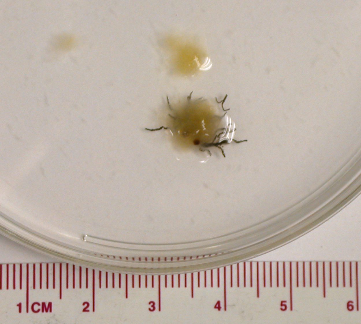
The following material was obtained from an ostomy bag.
Answer: not a parasite; most consistent with banana material. The twist to this case is that they are NOT actually banana seeds as is commonly taught in parasitology texts. Instead, they are polymerized tannins associated with xylem strands. This information was brought to my attention by Dr. Mary Parker, a microscopist at the Institute of Food Research, Norwich, UK (thank you Mary!). I therefore decided to show this case again so that everyone could benefit from this information.
Here is the full explanation:
The Cavendish bananas that are most often sold in grocery stores do not actually develop seeds. They are naturally sterile (triploid) and can only be propagated vegetatively. However, each aborted ovum has a vascular network consisting of xylem strands and associated cells containing astringent tannins. Upon ripening, the tannins polymerize into a semi-solid mass called 'tannin bodies' which fill the cells. The tannin bodies sometimes incorporate red-brown pigments from polyphenol oxidase activity (like the browning reaction in cut apples) as the cells age, and can therefore be seen as the red-brown bodies in this case. They are associated with the xylem strand which give them a chain-like appearance.
Because I've received some degree of skepticism when I've posted banana material in the past, I decided to conduct an experiment to see if I could recreate their appearance through some laboratory digestion techniques. So here was my process:
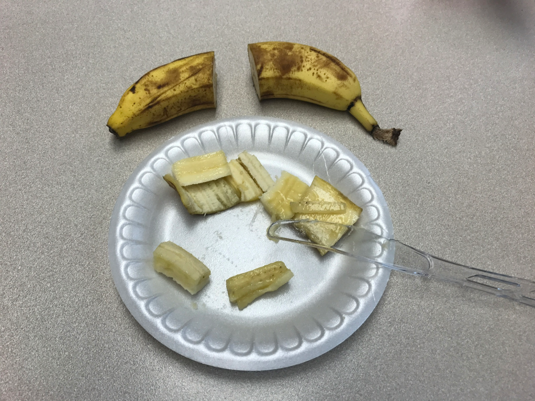
Step 1. Sacrifice my banana from lunch for the good of science
Note the darkly-staining structures that are seen in these longitudinal sections. These represent tannin bodies that have darkened over time.
Step 2. Add bananas to pre-prepared tubes of proteinase K in buffer. (Unfortunately I didn't have any amylase which would have digested the carbohydrates in the banana. However, this was the best I could do to simulate the digestive process). Vortex to mix and then incubate at 56 degrees Celsius while gently shaking (the standard tissue digestion that we use for PCR pre-processing).
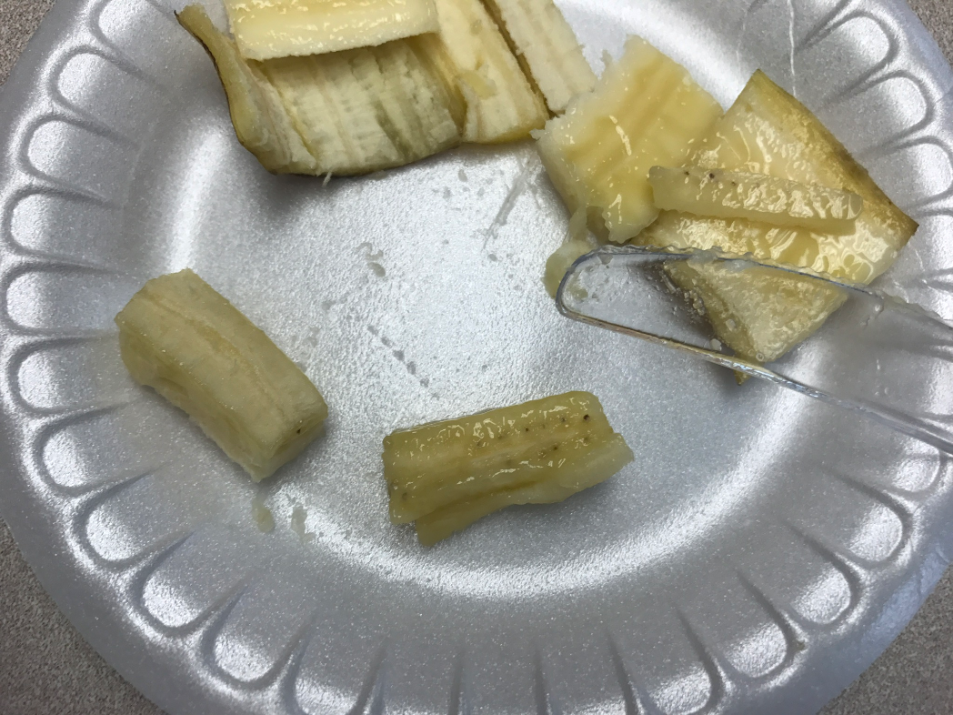
Step 3. Check regularly. I first checked every 10 minutes , but very quickly realized that this was going to be a long process. After the first 4 hours, this is how the banana sections looked:
Step 5. Final check - 48 hours. Success! I think that these look nearly identical to our clinical specimen. What do you think?
Close-up view of the tannin bodies and xylem strands (look a lot like the previous cases):
Again, it's not a perfect match since the actual patient specimen was subjected to the entire gastrointestinal digestive process. However, the strings of tannin bodies can clearly be seen. Here is how they look microscopically:
I hope you all enjoyed this experiment as much as I did!
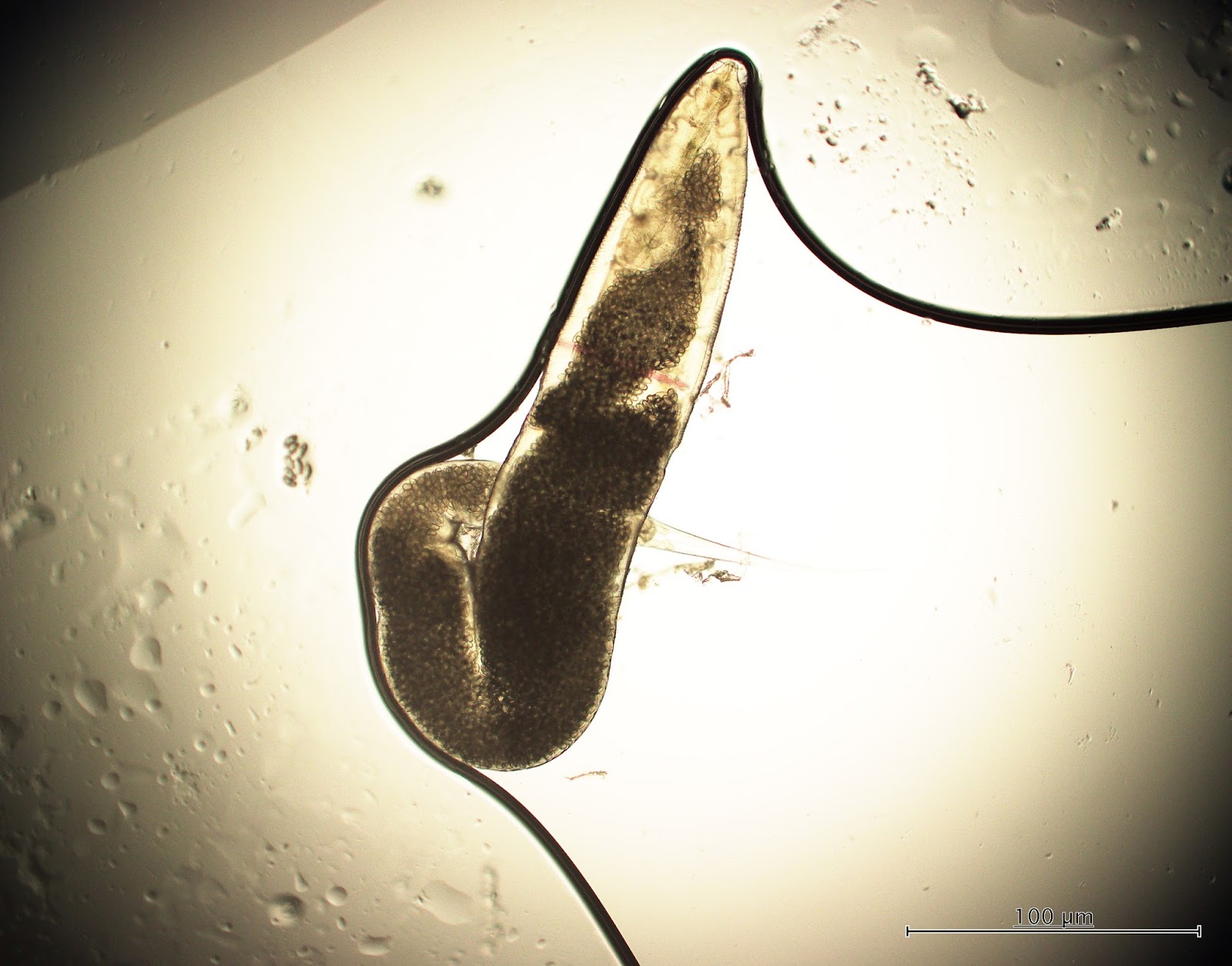
This week's post is the second in my new collaboration with the Institute of Tropical Medicine Antwerp and Idzi Potters. The following objects were seen in a urine specimen from a 73 year old man. They measure approximately 150 micrometers in length. The specimen was delayed in getting to the laboratory, and the patient indicated that it was mixed with toilet water.
Answer: Schistosoma sp. miracidium. Given the location in urine, the likely species is S. haematobium.
This case from Idzi Potters and the Institute of Tropical Medicine Antwerp shows the characteristic morphology of a form we rarely get to see in the laboratory - the motile ciliated miracidium that hatches from an egg when exposed to water. Note the interesting motility provided by the circumferential cilia.
You can recreate this in the lab by performing the 'hatching test' when you find Schistosoma eggs in stool or urine. There are instructions for the hatching test in most standard parasitology texts, although the process is somewhat time consuming.
In this case, the contamination of the urine specimen with toilet water and the delay in reaching the laboratory likely provided the stimulus and time needed for the eggs to hatch and release miracidia.
The primary differential diagnosis in this case is species of the free-living ciliate, Paramecium, that may be seen in fresh water. Idzi kindly provided the following videos of a Paramecium sp. so that we can appreciate the differences between them and schistosome miracidia. Paramecium spp. may range from 50 to 300 micrometers in length and therefore may overlap with the size range of S. haematobium miracidia (approximately 150 micrometers). They are also covered in circumferential cilia. The primary differences that we can appreciate at this magnification is that they are ovoid, lack an apical papilla (pointed apical end), and have rapid spiraling motility.
One reader suggested that the organisms seen in this case were miracidia from zoonotic schistosomes. This interesting suggestion prompted Idzi and I to do a little research! Idzi was able to find some very helpful studies to show that miracidia are unlikely to survive for more than 24 hours, and therefore couldn't have come from eggs that were passed by another animal and made it into the toilet water. According to Maldonado et al. (Biological studies on the miracidium of Schistosoma mansoni. Am J Trop Med 1948;28:645-657), the average life span of S. mansoni miracidia is 5 to 6 hours. Similarly, Lengy (Studies on Schistosoma bovis [Sonsino 1876] in Israel. Larval stages from egg to cercaria. Bull Res Counc Israel 1962;10:1-36) found that by 24 hours, all of the eggs in their study had hatched and all miracidia were dead. Ozgur Koru also noted that Schistosoma haematobium eggs will quickly hatch once being exposed to water (within 15 minutes). Based on these data, our conclusion is that the miracidia must have come from eggs in the patient's urine that hatched en route to the lab. Luckily the specimen reached the lab in time for the miracidia to be observed in their motile state.
Every week I will post a new Case, along with the answer to the previous case. Please feel free to write in with your answers, comments, and questions. Also check out my image archive website at http://parasitewonders.com. Enjoy!
The Fine Print: Please note that all opinions expressed here are mine and not my employer. Information provided is for educational purposes only. It is not intended as and does not substitute for medical advice. I do not accept medical consults from patients.