Sunday, October 30, 2016

Case of the Week 420

Happy Halloween! Here are some photos from my annual Halloween party - this year hosted with Dr. Audrey Schuetz and her husband Michael.

Our Clinical Microbiology fellow, Dr. Rachael Liesman. Can you guess which parasite she has?


Here are some yummy cupcakes that Rachael made:


Then there is our scary pizza delivery person, Heather:
And for extra bonus points, can you tell what parasite I am transmitting to this cat in my trail of slime? Hint: it's a cause of meningitis.

Saturday, October 29, 2016

Answer to Case 420

Answers:

Rachael is wearing a fish costume and is infected with an adult Diphyllobothrium latum, commonly known as the broad fish tapeworm. Yes, she technically would have the larval form rather than the adult worm, but hey, she gets extra bonus points for the excellent morphology of her worm. Note the 'spoon-shaped' scolex and the wide proglottids with central rosette-like uterine structures. Each proglottid was attached using Velcro, so she could 'shed' whole segments of proglottids at a time.

Her Giardia duodenalis cupcakes were delicious, just like her botfly and Dracunculus medinensis cupcakes from last year.

Heather delivered up Cryptosporidium pizza for all, along with her son who came dressed as a slice of pizza.

Lastly, you all guessed it - I was a snail shedding Angiostrongylus cantonenis larvae. Rats are the definitive host, whereas humans are accidental hosts and can get eosinophilic meningitis when accidentally ingesting larvae in infected snails, slugs, and slime. Cats aren't a known paratenic host, from what I could find in the literature, so my cat was probably fine.

Sunday, October 23, 2016

Case of the Week 419

This week's case was generously donated by Dr. Steve Jenkins. The object below was seen in a wet prep of semen. Identification?

Saturday, October 22, 2016

Answer to Case 419

Answer:  Ciliated epithelial cell. This cell likely arose from the caput epididymis, or possibly from an epididymal cyst of the male genitourinary tract that is lined by ciliated epithelium. The cilia continue to rhythmically beat even after the cells are detached from the rest of the epithelium (or even when the apical tuft containing the cilia has detached from the cell), and this may lead to a misdiagnosis of a parasitic infection. There are numerous reports of ciliated epithelial cells in respiratory epithelial cells being mistaken as trichomonads, free-living amebae, Balantidium coli, and even the insect commensal, Lophomonas. Less commonly, ciliated cells in male genitourinary specimens have been mistaken as parasites. Here is an older case report of a similar case that was mistaken for a parasite:
https://www.ncbi.nlm.nih.gov/pubmed/7495135
(email me if you want a copy of the article)

For future reference, here is a list of the sources of ciliated epithelium in the human body:
  • Upper respiratory tract
  • Fallopian tube and parts of the endometrium 
  • Ependymal cells that line the ventricles in the brain 
  • Caput epididymis/efferent ducts 
  • Any number of other locations where the epithelium has undergone a change called ciliary metaplasia (the epithelium converts from non-ciliated cells to ciliated cells)
Ciliated cells or detached ciliary tufts may be seen in specimens from any of these sources, and may be mistaken as parasites.

This was definitely a challenging case, and I loved seeing the great discussion that it generated! Huge kudos to Michigan Micro who pointed out the diagnostic features and led everyone away from a diagnosis of Trichomonas vaginalis (which was the predominant answer for quite a while). These key features are:

Reason #1. T. vaginalis doesn't have as many flagella as are seen in this case, and the flagella are more spaced out rather than grouped at one end. Compare the flagella of Trichomonas (top photo below, arrow) to the dense mat of cilia (arrow) on respiratory epithelial cells (bottom image):

Shown in the bottom image are detached ciliary tufts (left) and degenerated ciliated epithelial cells (right; arrow points to the apical portion of the cell where the cilia arise).

The bottom right image looks a lot like the following still photos taken from this case:


Reason #2: T. vaginalis has an undulating membrane, which is absent here (although it is hard to tell from the video).

Reason #3: T. vaginalis has 'jerky' motility (see my previous case of the week for trichomonad motility HERE - warning for obnoxious music)

Reason #4: T. vaginalis is usually more rounded or pear-shaped, whereas this specimen is more elongated.

The last component to consider is size, which unfortunately was not provided with the case. However, here are sizes of similar-appearing objects for comparison:
  • Trichomonas vaginalis trophozoites: 7-30 microns x 6-15 microns
  • Detached ciliary tufts: 10-15 microns diameter 
  • Intact ciliated columnar epithelial cells: Generally 4 times longer than wide, so this would be approximately 40-60 microns long x 10-15 microns wide.
  • Balantidium coli trophozoites: 40-100 microns in greatest dimension


Tuesday, October 18, 2016

Case of the Week 418

This week's case is from a young man from Africa with fevers. No further travel history is available. The following are images from peripheral blood preparations. Identification?

Unstained blood, Knott's concentration, 10x objective (100x total magnification):

Unstained blood, 40x (400x total):

Thick film, Giemsa, 50x (500x total):


Thick film, Giemsa, 100x (1000x total):


Sunday, October 16, 2016

Answer to Case 418

Answer: Loa loa

Florida Fan nicely describes the important features of this case:
"The microfilariae are sheathed which narrows the choice to only three: Wuchereria bancrofti, Loa loa and Brugia malayi/Brugia timori. The sheath did not stain pink, and the patient did not come from Southeast Asia or Polynesia, thus eliminating B. malayi" (although the pink sheath is not always seen with B. malayi). "The column of nuclei extends all the way to the end of the tail and this differentiates these microfilariae from those of W. bancrofti. We have one last choice: Loa loa." 

Blaine will remind us that the sheath is not always seen, and therefore the size of the microfilariae should also be taken into consideration. The unsheathed microfilariae of Mansonella spp. are significantly narrower than the others, which is a helpful feature in eliminating this filarial worm from the differential.

Here are some of the key diagnostic findings seen in the images in this case:


Tuesday, October 11, 2016

Case of the Week 417

This week's case is a cool little finding from my lab, with videos by Emily F. Specimen type is urine (filtered through a Nuclepore (R) membrane), and the object shown measures approximately 150 micrometers in length.


Identification?

Monday, October 10, 2016

Answer to Case 417

Answer: Schistosoma haematobium egg containing a live miracidium.

The video in this case nicely shows the movement of the miracidium as it beats its circumferential cilia and retracts its apical papilla. Normally internal "flame" cells (protonephridium) can also been seen moving, but these cannot be appreciated in this video.


Wednesday, October 5, 2016

Case of the Week 416

This week's case was generously donated by Mr. Boren Huot, and is in honor of George from Memorial Sloan Kettering Cancer Center, who just showed me a very striking example of a similar case.

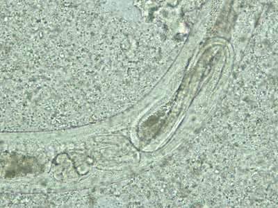
The patient is a young woman with watery diarrhea. The following organisms were seen in stool concentrate. Identification?


Tuesday, October 4, 2016

Answer to Case 416

Answer: Strongyloides stercoralis adult female worms containing eggs and rhabditiform larvae. A possible filariform larva is also seen.

Adults worms are almost never seen in the stool of patients with strongyloidiasis, but when present, indicate a likely heavy infection. This case is remarkable because eggs and larvae are seen within the adult worms, illustrating how quickly the eggs hatch in the normal life cycle of this worm. The adult female usually lays the eggs directly in the intestinal mucosa where they quickly hatch to release rhabditiform larvae. This is why larvae, and not eggs, are found in the stool of patients with S. stercoralis infection.

Thanks again to Boren for donating this fascinating case.