Saturday, July 30, 2016

Case of the Week 407

This week's amazing case was donated by Dr. Sue Whittier. The patient had cooked salmon and eaten it with her family. It was then refrigerated and reheated it for dinner the next day. Here is what the diners saw in the re-heated salmon:

It definitely caught everyone's attention since it was actively moving!

 Identification? What follow-up would you recommend for the individuals who had eaten this salmon the day before?

Friday, July 29, 2016

Answer to Case 407

Answer: anisakid larva, most likely Pseudoterranova species.

This case is a dramatic example of what you can find if you don't cook your fresh, unfrozen salmon before eating it! Anisakids (Anisakis spp. and Pseudoterranova spp.) are very common in wild-caught salmon, cod, and other fish, and therefore, fish should be fully cooked or frozen prior to being ingested. Freezing at -20C for 24 hours would probably kill any anisakids, but the trematodes and cestodes are a bit more resilient. Therefore, the FDA recommends freezing fish at -20C for 7 days, or -70C for 24 hours before consuming it raw or undercooked.

Differentiation of Anisakis from 
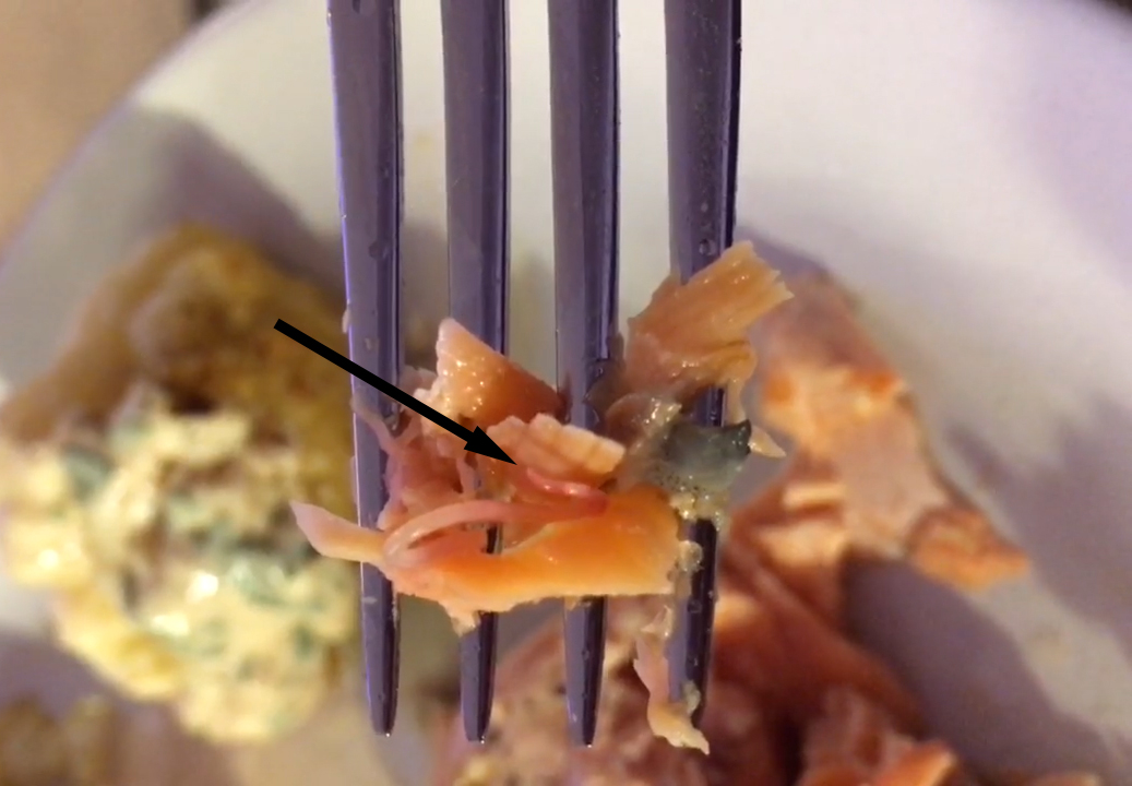
Pseudoterranova usually requires close examination of the larva, looking for the cecum. However, Blaine kindly pointed out that you can actually catch a glimpse of what is most likely the cecum in the video I provided. Here is a still image from the video:
The arrow points to the light tan structure which is most likely the cecum. 

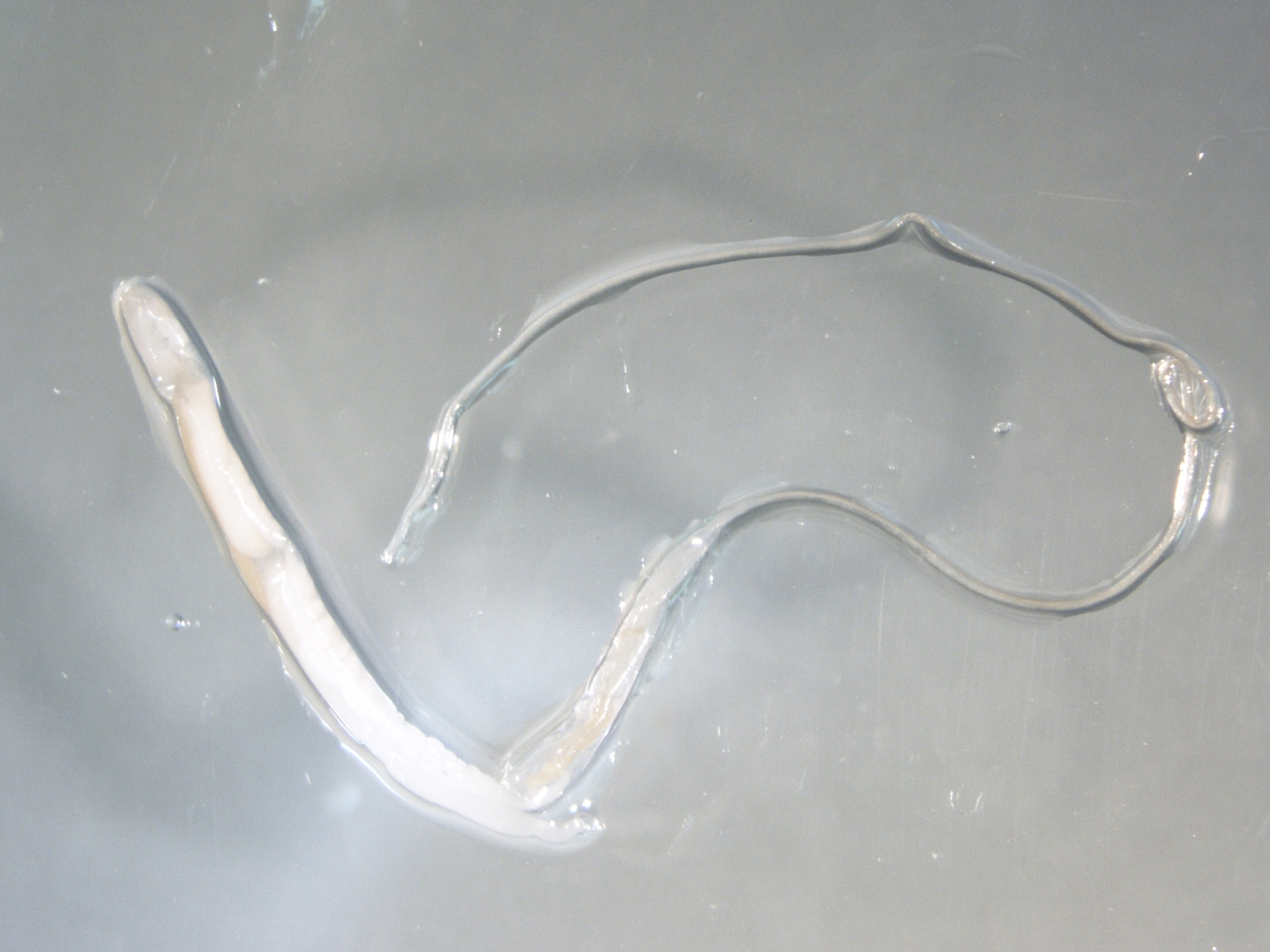
Here is an image of a live Pseudoterranova worm from the CDC DPDx group for comparison (arrow points to the cecum):

To answer the question about patient management - no treatment is necessary unless the patient is symptomatic, at which time albendazole and/or endoscopic examination to remove an embedded worm may be needed. Prophylactic albendazole could also be given, but there are no clear-cut recommendations on this. 

Tuesday, July 26, 2016

Case of the Week 406

This week's case was generously donated by Dr. Kamran Kadkhoda. This arthropod was found on a young girl near Winnipeg, in Manitoba, Canada.


Identification?

Sunday, July 24, 2016

Answer to Case 406

Answer: Dermacentor sp. tick, adult male

Wow, lots of great discussion on this case! Thank you readers for providing the identification keys and references. Some of the key features that identify this as a Dermacentor are the ornate scutum, short mouthparts, and festoons. Examination of the spiracular plates is necessary for differentiation between D. variabilis and D. andersoni, and, as Jon mentioned, you can't see the spiracular plates well enough in these photos to make that distinction.


I believe that the CDC pictorial keys that Florida Fan was referring are HERE. These are definitely a good place to start when performing arthropod ID. I'll also put in a shameless plug for a benchtop reference guide that Blaine Mathison and I published with the CAP Press that provides guidance for identification of arthropods to the genus level (which we don't make money on, so I don't feel bad mentioning). Finally, Blaine and I published a review on identification of medically-important arthropods through Clinical Microbiology Reviews that you can access HERE. If you don't have access to the full text, send me an email at b_pritt@yahoo.com and I will send you a copy.

Monday, July 18, 2016

Case of the Week 405

This week's case is from my own lab, with the beautiful images taken by Emily F. The patient is an asymptomatic male who underwent routine screening colonoscopy, which revealed the following:
Two objects were removed during colonoscopy and submitted to the Clinical Parasitology lab for identification:

 By manipulating the objects, we were able to express the following:


Identification?

Sunday, July 17, 2016

Answer to Case 405

Answer: Trichuris trichiura (whipworm)

As pointed out by Arthur V and the other readers, the shape of the adult worms (narrow anterior end, broad posterior end) and the eggs are characteristic for this roundworm. Note that there is both a female (second image, straight posterior) and a male (image, curved posterior) in this specimen.

Another interesting feature of this case is that we were able to express both immature and mature eggs out of the female worm using blunt manipulation. The differences in maturity are responsible for the slightly different appearances of the eggs (kudos to the readers that pointed this out!) Here are the 2 eggs side-by-side for comparison:


Sunday, July 10, 2016

Case of the Week 404

This week's case was generously donated by Florida Fan. This structure was found in a concentrated stool specimen from a teenage male. It measures approximately 45 micrometers in diameter (shown at 400x total magnification). Identification?


Saturday, July 9, 2016

Answer to Case 404

Answer: Hymenolepis nana egg

As pointed out by Sheldon, BW, and Anon, this is a beautiful example of an H. nana egg, with refractile internal hooklets and 2 clearly visible polar thickenings. The arrows on the image below point out the polar thickening from which the polar filaments arise.

Thanks again for Florida Fan for donating this great case!

Sunday, July 3, 2016

Case of the Week 403

Happy Independence day to all of my American readers! Here is some red (magenta), white and blue for you - found in a modified acid fast-stained stool specimen:


 The objects measure approximately 35 micrometers in length. Identification?

Saturday, July 2, 2016

Answer to Case 403

Answer: Cystoisospora (formerly Isospora) belli

As pointed out by the readers, these are immature oocysts, since only 1 sporoblast is visible.

This is the state that this organism is shed in the stool of infected individuals. The oocysts then mature in the environment into their infective form, with the sporoblast dividing into 2 sporoblasts. The sporoblasts develop cell walls and become sporocysts containing 4 infective sporozoites each.

Thanks to Florida Fan for pointing out that the modified/hot safranin stain will also beautifully stain the oocysts of Cystoisospora (and other coccidia). The oocysts also exhibit striking autofluorescence using UV microscopy (excitation filter at 330-365 nm or 450-490 nm).

I hope you all had a safe and happy July 4th weekend!