This week's case was generously donated by Dr. Rocco LaSala. The specimen was retrieved from the stool of a 70 year-old-woman with approximately 12 months of diarrhea, abdominal cramping, and unintended weight loss (> 40 lbs). The structures are thin, brown, and 'thread-like', measuring approximately 0.7 mm in width.
Specimens were also submitted for histology, which showed the following on H&E staining:
This case was clearly not a parasite (or even animal tissue), as seen on both gross and microscopic images. The material most closely resembles banana seeds, as seen on a previous case that I've posted.
Unfortunately, this doesn't help elucidate the cause of the patient's symptoms, and therefore additional studies will be necessary.
Thanks again to Rocco for sharing this case with us.
Answer: Dermacentor sp. ticks: an adult male and female (latter is heavily engorged) and newly-laid eggs.
We were very excited to find these eggs in the container along with the live female. Therefore, we decided to try to hatch them. Special thanks to William Nicholson and Graham Hickling who provided the necessary instructions, and to Jim, Lynn, Heather, Emily and the rest of my awesome lab staff who made it all happen. It turns out that hatching larvae from eggs is not a straight-forward process. The eggs need to be maintained around room temperature, and importantly, need to be kept in a humid environment (e.g. 80-90%). Also, they need to be monitored regularly for fungal growth which could kill them (I can provide instructions to anyone who is interested). To accomplish this, we set up the following humidity chamber:
We went through a couple of different iterations of the humidity chamber, using different solutions and containers, but eventually settled on the design shown here - a simple sealed Tupperware container with distilled water at the base. The tick eggs are in a Petri dish sealed with gas-permeable tape and elevated above the water on a used pipette tip holder).
We then patiently waited for several weeks, monitoring the eggs closely.
As a reminder, here is what the eggs shortly after being laid (May 20):
We were excited to see that they appeared to mature after ~4.5 weeks of incubation (June 17):
By June 23rd, they looked ready to hatch:
And sure enough, by the next day we had hatching larvae!
So now you may be wondering what we are going to do with all of them? Well, as cute as they are, they're still baby ticks that could be harboring human pathogens, so they will be dumped into formalin to be preserved for posterity.
Welcome to our 400th Parasite Case of the Week! To celebrate the 400th case, I thought I would dedicate this post to education in parasitology and share with you 5 of my favorite parasite teaching tools.
1. Embed arthropods and worms in casting resin, using products purchased at your local arts and crafts store. I've used Castin' Craft in the past, but there are other options out there as well. Here is one of my creations - adult Ixodes scapularis ticks (in different stages of engorgement) in a small petri dish:
Embedded arthropods are great for teaching because they are durable (e.g. their legs don't fall off from being manipulated over and over again), and you can easily examine both their dorsal and ventral aspects. If you're feeling really creative, then you can also make them into little works of art - or even jewelry (start a fun new trend). You can buy molds in every shape and size under the sun.
2. Find some anisakids in frozen (or fresh) cod. I have the best luck with Atlantic cod; in every bag of 10 fillets, I usually find at least 1 worm. I let the fillets thaw in the refrigerator and then use blunt dissection to find the coiled larvae. I put the fish (with anisakid) on a plate for display in my teaching lab. It's a great way to get medical students, residents, techs, and clinicians interested in parasitology.
For added fun, have a fish dissecting party! Here are some of my awesome pathology residents, who came up with this idea all on their own.
3. Buy some medicinal leeches (Hirudo medicinalis) at your hospital pharmacy. Like the cod, these also make a great display for a parasite teaching laboratory. You can use them as an example of an ectoparasite, talk about the anticoagulant they produce (hirudin - used medicinally), discuss their fascinating history (past and present) in medicine, and also discuss why it is necessary to give prophylactic antibiotics to recipients of leech therapy (to prevent sepsis with Aeromonas - a commensal bacterium in the gut of medicinal leeches).
Yes, it's a display with shameless 'ick' factor, but it's great for capturing the attention of medical students and clinicians alike.
4. Learn some basic entomology skills. Try dragging for ticks, collecting black fly (Simulium sp.) larvae from a fast-flowing stream (below), or learn how to mount insects professionally for teaching purposes (compliments of Blaine Mathison).
5. Last, but not least, have a parasite potluck, with treats decorated with your favorite (candy) parasites.
Cupcakes by Dr. Rachael Liesman, my Clinical Microbiology fellow:
Malaria cookies by my Education Specialist, Emily Fernholz
Cakes from the Mayo Medical Students! Coconut rectum, Giardia duodenalis, Reduviid bug transmitting Trypanosoma cruzi through its feces, Strongyloides stercoralis on a stool agar culture, and a strawberry cervix.
So for my questions this week: What parasites are associated with a 'coconut rectum' and 'strawberry cervix'?
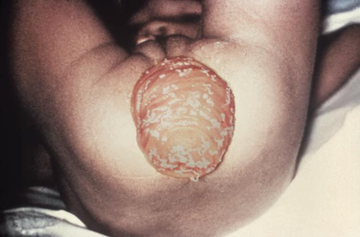
1. Parasite associated with 'coconut rectum' - Trichuris trichiura. This description refers to a prolapsed rectum on which numerous adult worms attached to the rectal mucosa can be seen.
(Image courtesy of the CDC Public Health Image Library)
2. Parasite associated with 'strawberry cervix' - Trichomonas vaginalis. This description refers to the punctate hemorrhages seen on the infected cervix which purportedly look like a strawberry.
(Image courtesy of the CDC Public Health Image Library)
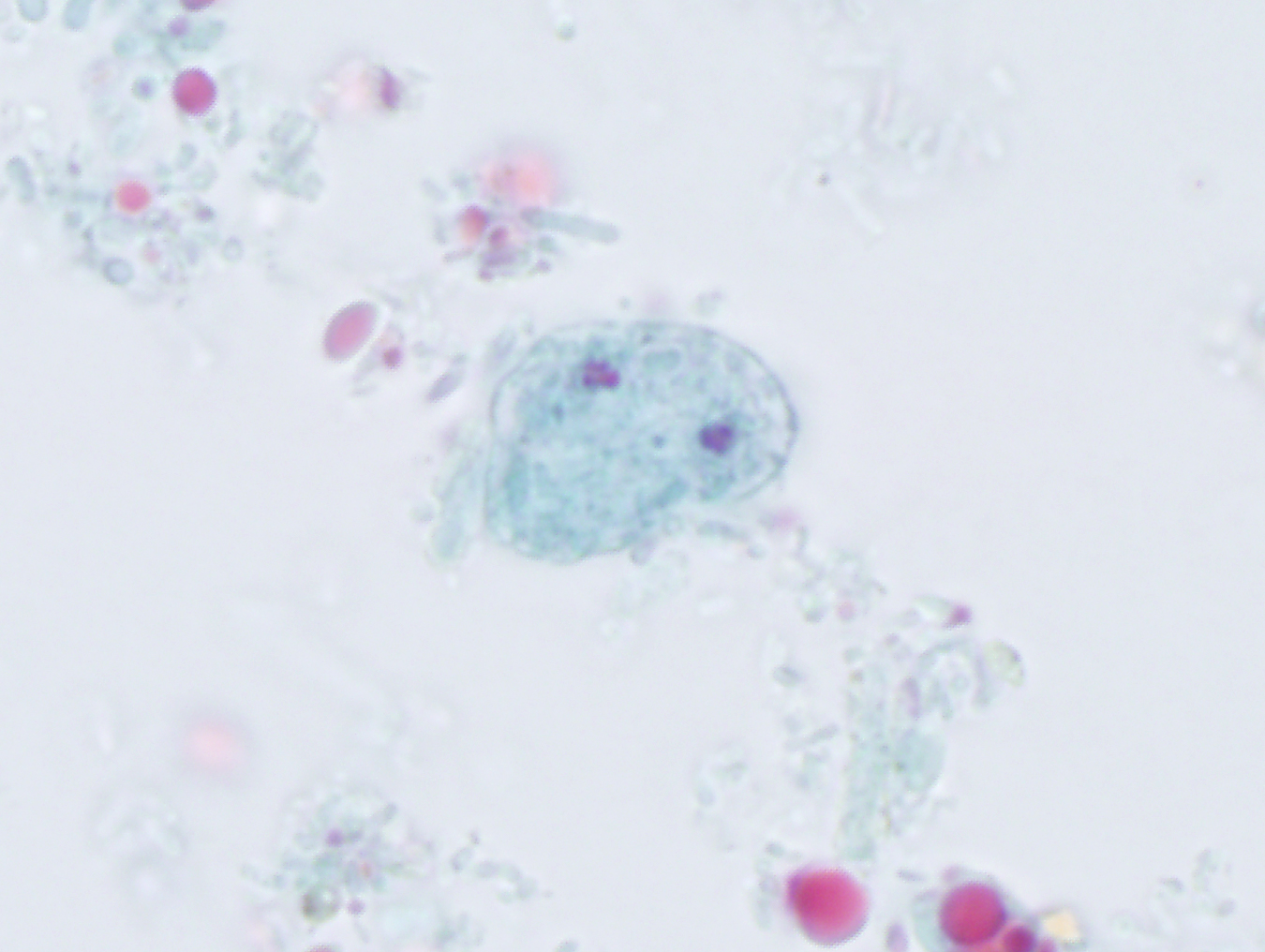
Wow, we're almost at 400 cases! I will have to think of something extra special for next week. Meanwhile, here is classic finding in a trichrome-stained stool specimen (1000x).
Answer: Dientamoeba fragilis
This organism has an overall structure like an amoeba, but has an internalized flagellum, and is therefore most accurately characterized as a flagellate. It is sometimes referred to as an amoeboflagellate. As nicely described by Florida Fan, D. fragilis has a characteristic nucleus with 'fragmented' appearing chromatin. It commonly has 2 nuclei, but may also have 1, as seen in some of the organisms in this case.
Every week I will post a new Case, along with the answer to the previous case. Please feel free to write in with your answers, comments, and questions. Also check out my image archive website at http://parasitewonders.com. Enjoy!
The Fine Print: Please note that all opinions expressed here are mine and not my employer. Information provided is for educational purposes only. It is not intended as and does not substitute for medical advice. I do not accept medical consults from patients.