Tuesday, December 27, 2016

Case of the Week 427

Welcome back for the last case of 2016!

I thought I would close out the year with an old favorite. The case and beautiful images are donated from George at MSKCC. The specimen is concentrated stool from a young child.


Identification of the 2 worms shown here?

Monday, December 26, 2016

Answer to Case 427

Answer: Enterobius vermicularis (pinworm) male and female adults

The adult worms are easily identified by their prominent lateral alae:

The male and female can be further differentiated by looking at their tails; males have a coiled tail while females have a straight tail.

This case is unusual for a couple of reasons; first, we don't typically see adult pinworms in stool specimens (they are more commonly [and ideally] detected using tape preparations), and second, it is even less common to see male pinworms in clinical specimens. The females are more commonly seen because they are the ones that leave their safe residence in the large intestine/cecum to travel nightly to the perianal folds to lay their eggs. The males typically remain behind. The fact that we see both male and female pinworms in stool makes me wonder if this was a particularly heavy infection.

Thank you again to George from MSKCC for donating this interesting case!

Sunday, December 18, 2016

Case of the Week 426

Happy Holidays to all of my readers! Can anyone tell me who the happy couple is below the mistletoe?


Saturday, December 17, 2016

Answer to Case 426

Answer: Schistosoma sp. male and female couple "in copula"

The adult female of this trematode is smaller than the male and can generally be found resting in the copulatory groove of the larger male.

Wednesday, December 7, 2016

Case of the Week 425

This week's case was generously donated by Dr. Mike Mitchell. The patient is a teenage girl with vague abdominal complaints. No anal pruritus was noted. The following were seen on colonoscopy (cecum).

A biopsy was also performed which showed the following:


Identification?

Tuesday, December 6, 2016

Answer to Case 425

Answer: Enterobius vermicularis

This was a challenging case, since we aren't given the size of the worms, their movement in the video precluded close examination, and the histologic features were somewhat distorted.

While I had initially thought of E. vermicularis, I must admit that, with further evaluation, I had convinced myself that the "anterior end" was embedded into the intestinal mucosa, making me instead suspect Trichuris trichiura (arrow below)
Thankfully, my parasite pals, Blaine Mathison and Dr. Marc Courturier, provided a valuable second opinion. After our discussion, I realized that the end shown above was actually the partially clear posterior that we often see with female pinworms, like in the image below (arrow):
Blaine also pointed out that the tall musculature seen with T. trichiura was lacking in the biopsy specimen, thus ruling out that diagnosis. Also, he noted that he has occasionally seen the lateral alae take on a similarly distorted appearance in biopsy specimens: 
Therefore, the lessons for me on this case were 1.) it's possible to "overthink" yourself out of the correct diagnosis and 2.) reach out to your parasite pals when you are in need of a second opinion. 
We have built a strong community of parasitologists here through this blog, and I thank all of you for your ongoing support. Happy Holidays!

Sunday, November 27, 2016

Case of the Week 424

The following are Giemsa-stained peripheral blood smears from a young girl who was recently treated for malaria. What is the material inside of the white blood cells?


Saturday, November 26, 2016

Answer to Case 424

Answer: hemozoin pigment

This pigment is formed as the infecting Plasmodium species feeds on the host's hemoglobin in the red blood cells. This young girl had previously been diagnosed with a heavy Plasmodium falciparum infection (3.5% parasitemia). Here are some images of her previous thick and thin blood films.

Fortunately, she made a full recovery. Thank you again to Emily F. for these beautiful photos.

Monday, November 21, 2016

Case of the Week 423

The following objects were observed in an aspirate of liver cyst fluid (carmine stain). Identification?

40x

100x

200x

400x

Sunday, November 20, 2016

Answer to Case 423

Answer: Echinococcus sp. protoscoleces

Each protoscolex has an internal row of hooklets that will evert one inside of the canine definitive host and become the head of the adult tapeworm.

A couple of readers asked me about the Carmine stain. It's unfortunately not one that we perform in the lab anymore although there are instructions and recipes in many major parasitology texts and online. It's really a beautiful way to highlight the structures of various worms, but just not something that we usually need for diagnostic purposes. I try to show photos of these 'older' stains from time to time to hopefully inspire others who want to revive them for teaching purposes.

Sunday, November 13, 2016

Case of the Week 422

Here is another lovely photograph from Florida Fan - a straight forward ID for those of you studying for boards or your Trop Med exam.

This object was observed in a stool specimen from a middle-aged man from India. It measures approximately 30 microns in diameter.

Identification?

Saturday, November 12, 2016

Answer to Case 422

Answer: Taenia species, egg

As Deepak described, the characteristic features include the small size, yellow-brown color and thick radially striated shell. Note the presence of a six-hooked embryo (oncosphere) inside.

Unfortunately you can't tell the different Taenia species (i.e. Taenia solium, T. saginata, T. asiatica) apart from the egg alone. This would require molecular testing and/or examination of the scolex or proglottid morphology.

Thanks again to Florida Fan for donating this classic example of a Taenia eggs.

Monday, November 7, 2016

Case of the Week 421

This week's beautiful photographs came from Florida Fan. The specimen came from a middle-aged male.

By spinning down the 70% ethanol that the specimen had been transported in, the following objects were seen. They measured approximately 60 micrometers in length.

Identification?

Sunday, November 6, 2016

Answer to Case 421

Answer: Diphyllobothrium species
There are several characteristic morphologic features shown in these nice images from Florida Fan. First, we have a long segment of a relatively large tapeworm, with proglottids that are wider than they are long. Immature proglottids of Taenia solium and T. saginata can also be wider than they are long, but the fact that we could get eggs from the proglottids means that they are mature. Therefore, we can exclude Taenia based on the shape of the proglottids. We can also exclude the other human tapeworms since none are this large.

Another feature that I find to be helpful for identifying Diphyllobothrium is that the uterine structures are clearly visible in the center of each proglottid - even without staining or microscopic examination:

Finally, the eggs that were found in the accompanying fluid leave no doubt as to the identification, having the classic shape and features of Diphyllobothrium, including an operculum and abopercular knob.

As Anon pointed out, species identification would require epidemiological information, and ultimately molecular testing. Therefore, we report this out as "Diphyllobothrium species" in my lab.

Sunday, October 30, 2016

Case of the Week 420

Happy Halloween! Here are some photos from my annual Halloween party - this year hosted with Dr. Audrey Schuetz and her husband Michael.

Our Clinical Microbiology fellow, Dr. Rachael Liesman. Can you guess which parasite she has?


Here are some yummy cupcakes that Rachael made:


Then there is our scary pizza delivery person, Heather:
And for extra bonus points, can you tell what parasite I am transmitting to this cat in my trail of slime? Hint: it's a cause of meningitis.

Saturday, October 29, 2016

Answer to Case 420

Answers:

Rachael is wearing a fish costume and is infected with an adult Diphyllobothrium latum, commonly known as the broad fish tapeworm. Yes, she technically would have the larval form rather than the adult worm, but hey, she gets extra bonus points for the excellent morphology of her worm. Note the 'spoon-shaped' scolex and the wide proglottids with central rosette-like uterine structures. Each proglottid was attached using Velcro, so she could 'shed' whole segments of proglottids at a time.

Her Giardia duodenalis cupcakes were delicious, just like her botfly and Dracunculus medinensis cupcakes from last year.

Heather delivered up Cryptosporidium pizza for all, along with her son who came dressed as a slice of pizza.

Lastly, you all guessed it - I was a snail shedding Angiostrongylus cantonenis larvae. Rats are the definitive host, whereas humans are accidental hosts and can get eosinophilic meningitis when accidentally ingesting larvae in infected snails, slugs, and slime. Cats aren't a known paratenic host, from what I could find in the literature, so my cat was probably fine.

Sunday, October 23, 2016

Case of the Week 419

This week's case was generously donated by Dr. Steve Jenkins. The object below was seen in a wet prep of semen. Identification?

Saturday, October 22, 2016

Answer to Case 419

Answer:  Ciliated epithelial cell. This cell likely arose from the caput epididymis, or possibly from an epididymal cyst of the male genitourinary tract that is lined by ciliated epithelium. The cilia continue to rhythmically beat even after the cells are detached from the rest of the epithelium (or even when the apical tuft containing the cilia has detached from the cell), and this may lead to a misdiagnosis of a parasitic infection. There are numerous reports of ciliated epithelial cells in respiratory epithelial cells being mistaken as trichomonads, free-living amebae, Balantidium coli, and even the insect commensal, Lophomonas. Less commonly, ciliated cells in male genitourinary specimens have been mistaken as parasites. Here is an older case report of a similar case that was mistaken for a parasite:
https://www.ncbi.nlm.nih.gov/pubmed/7495135
(email me if you want a copy of the article)

For future reference, here is a list of the sources of ciliated epithelium in the human body:
  • Upper respiratory tract
  • Fallopian tube and parts of the endometrium 
  • Ependymal cells that line the ventricles in the brain 
  • Caput epididymis/efferent ducts 
  • Any number of other locations where the epithelium has undergone a change called ciliary metaplasia (the epithelium converts from non-ciliated cells to ciliated cells)
Ciliated cells or detached ciliary tufts may be seen in specimens from any of these sources, and may be mistaken as parasites.

This was definitely a challenging case, and I loved seeing the great discussion that it generated! Huge kudos to Michigan Micro who pointed out the diagnostic features and led everyone away from a diagnosis of Trichomonas vaginalis (which was the predominant answer for quite a while). These key features are:

Reason #1. T. vaginalis doesn't have as many flagella as are seen in this case, and the flagella are more spaced out rather than grouped at one end. Compare the flagella of 
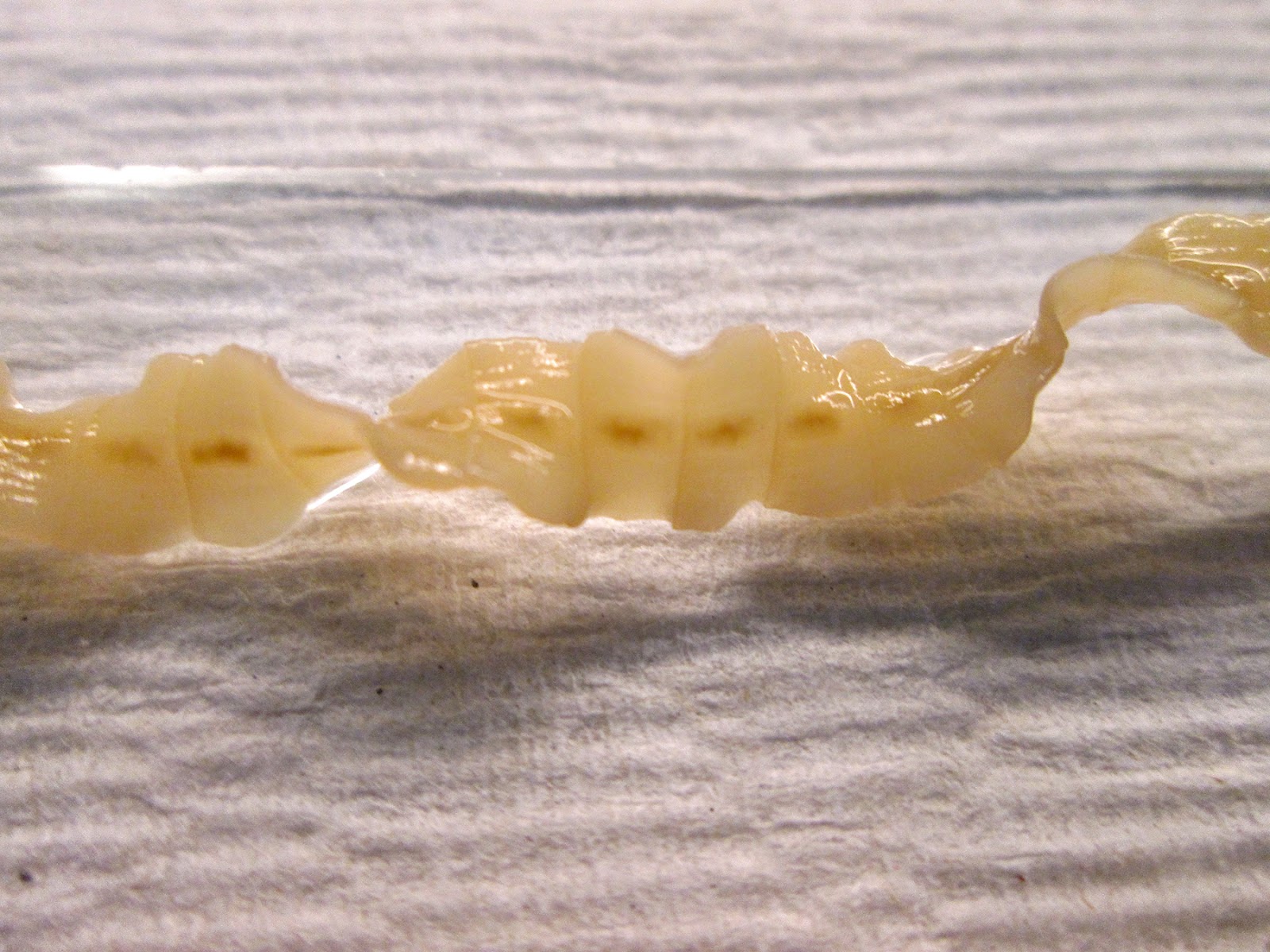
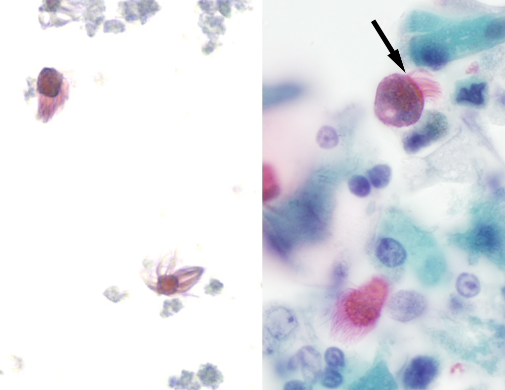
Trichomonas (top photo below, arrow) to the dense mat of cilia (arrow) on respiratory epithelial cells (bottom image):

Shown in the bottom image are detached ciliary tufts (left) and degenerated ciliated epithelial cells (right; arrow points to the apical portion of the cell where the cilia arise).

The bottom right image looks a lot like the following still photos taken from this case:


Reason #2: T. vaginalis has an undulating membrane, which is absent here (although it is hard to tell from the video).

Reason #3: T. vaginalis has 'jerky' motility (see my previous case of the week for trichomonad motility HERE - warning for obnoxious music)

Reason #4: T. vaginalis is usually more rounded or pear-shaped, whereas this specimen is more elongated.

The last component to consider is size, which unfortunately was not provided with the case. However, here are sizes of similar-appearing objects for comparison:
  • Trichomonas vaginalis trophozoites: 7-30 microns x 6-15 microns
  • Detached ciliary tufts: 10-15 microns diameter 
  • Intact ciliated columnar epithelial cells: Generally 4 times longer than wide, so this would be approximately 40-60 microns long x 10-15 microns wide.
  • Balantidium coli trophozoites: 40-100 microns in greatest dimension


Tuesday, October 18, 2016

Case of the Week 418

This week's case is from a young man from Africa with fevers. No further travel history is available. The following are images from peripheral blood preparations. Identification?

Unstained blood, Knott's concentration, 10x objective (100x total magnification):

Unstained blood, 40x (400x total):

Thick film, Giemsa, 50x (500x total):


Thick film, Giemsa, 100x (1000x total):