Monday, March 30, 2015

Case of the Week 343

The following biopsies of small intestine are from a patient with chronic watery diarrhea.  Diagnosis? (CLICK ON IMAGES TO ENLARGE).

H&E, 200x

H&E, 400x

H&E, 1000x

I'd like to thank Dr. Duane Newton who allowed me to borrow his slides and take these photographs.

Sunday, March 29, 2015

Answer to Case 343

Answer:  Cyclosporiasis due to Cyclospora cayetanensis

Note the presence of both asexual and sexual forms within enterocytes:

As many readers noted, the differential diagnosis includes Cryptosporidium sp., Cystoisospora belli, and the microsporidia - all which can also be found within enterocytes.  They can be differentiated from Cyclospora cayetanensi by their size and location within the cell.

Cryptosporidium spp. undergo a complicated replication cycle outside of the enterocyte cytoplasm, just below the cell membrane (thus they are intra-cellular, extra-cytoplasmic).  They therefore appear as small 'blobs' on the surface of the intestine (arrow heads below):

Cystoisospora belli is more like Cyclospora cayetanensis in that the parasites replicate within the cytoplasm of the enterocytes. They are significantly larger than Cyclospora, which in my mind, is the main differentiating feature (images from the CDC DPDx):

Finally there are the microsporidia (usually Enterocytozoon bieneusi and Encephalitozoon intestinalis), which appear as very small intra-cytoplasmic objects.  You can see a nice Giemsa-stained image from Para-site online.

Sunday, March 22, 2015

Case of the Week 342

A 50-year old man presented with peritonitis and an acute abdomen. Emergent surgery revealed a perforation of his small intestine. Further history revealed that he had recently traveled to Costa rica.

The following are H&E stained sections of his small intestine showing unexpected structures in a blood vessel with extensive surrounding eosinophilic inflammation.

20x total magnification

40x total magnification
 200x magnification


 Around the worm were numerous eosinophils and granulomas containing foreign objects.  Here is one such granuloma.

Identification?

Saturday, March 21, 2015

Answer to Case 342

Answer: Angiostrongylus costaricensis

This is an interesting case in that we were able to see nice longitudinal and cross-sections of the adult worm(s) within a blood vessel in the wall of the small intestine. The morphologic features, including intestine and (somewhat compressed) lateral cords are clearly visible. Foreign bodies surrounded by a granulomatous response are also seen in the tissue around the worm and most likely represent eggs or larvae.  The fact that the patient had recently travelled to Costa Rica fits perfectly with the identification and supports our morphologic diagnosis.  Here are some of the characteristic features:

Adult female:
Possible egg (arrow) within a granuloma. Note the abundant eosinophilic host response.
John B. suggested the possibility of a that there is a protist within this nematode.  A great thought, although I believe what we are seeing are just the internal structures of A. costaricensis. Thank you all for the great comments!

Monday, March 16, 2015

Case of the Week 341

The following images are from an autopsy of a woman with end-stage ovarian cancer who developed respiratory failure and sepsis.  The tissue type is small intestine. This is an old case from the Armed Forces Institute of Pathology (AFIP) that I found in my files over the weekend.  It is such a shame that they closed - they were such a wonderful resource.

Questions:
1. What is the diagnosis?
2. What forms of the parasite are shown in these images


?


Sunday, March 15, 2015

Answer to Case 341

Answer:  Strongyloidiasis due to Strongyloides stercoralis

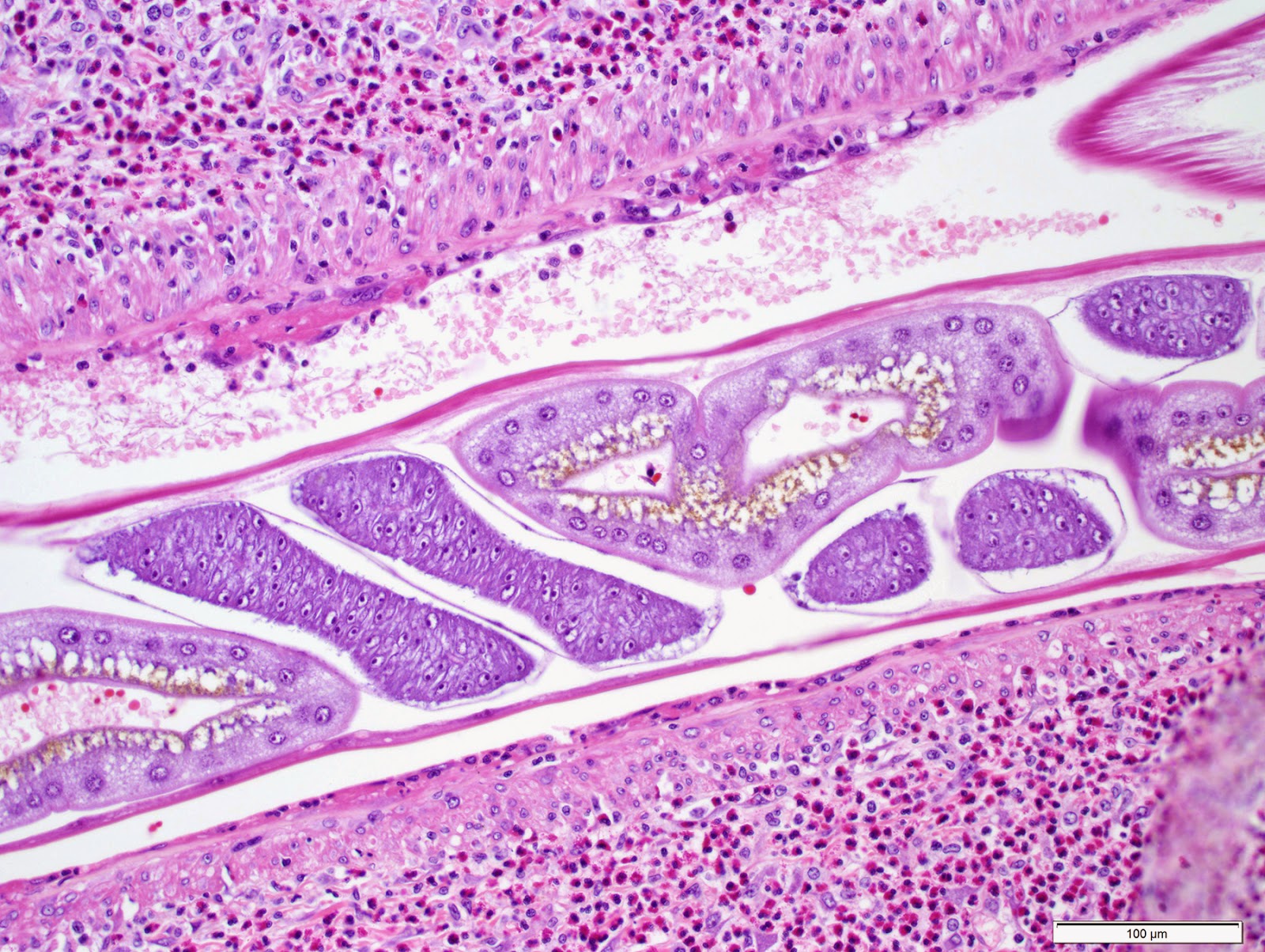
As with most biopsies of S. stercoralis, all 3 parasite forms can be seen:  the adult female, larvae and eggs:

Note that the eggs resemble those of hookworm.  However, unlike human hookworm infection, eggs are never seen in the stool since they hatch to release rhabditiform larvae within the intestine.  I like how you can see different stages of cell division within the eggs in this case:


As mentioned by Arthur, this diagnosis fits with the patient's clinical presentation. He states "I would guess that it (the infection) was benign until immunosuppressive chemotherapy began to treat the cancer, at which point a hyperinfective syndrome developed due to the opportunistic and autoinfective nature of this worm. The respiratory failure is likely due to the damage the worms do during the pulmonary stages of the life cycle, and sepsis is well documented in hyperinfective strongyloidiasis due to the filariform larvae penetrating the mucosa to reach the circulatory system, dragging coliform bacteria into the blood stream."

Of note, we take advantage of the propensity for larvae to drag bacteria with them when we use the stool agar culture method to detect Strongyloides stercoralis and other nematodes.  When present in a stool specimen, the larvae migrate into the surrounding agar, dragging bacteria with them. The bacteria grow in the tracks, thus producing visible evidence of the larval migration. The agar culture method is the most sensitive means for detecting Strongyloides stercoralis in stool (slightly more sensitive than the older Baermann method - and nicer to perform from a laboratory standpoint because there is no stool in an open container on the benchtop!)  You can see some images of S. stercoralis on culture plates in Case of the Week 137, and most recently, by the impressive case donated by Lee (Case of the Week 337). Note that you don't need a special type of agar to see the tracks left by Strongyloides - anything that supports bacterial growth will work. The risk comes from accidental exposure to laboratory techs when they handle the plates with bare hands since Strongyloides stercoralis and hookworm larvae can penetrate intact skin. When we perform the agar culture test in the parasitology lab at my institution, we always tape up the plates and put them in a transparent plastic bag for incubation at room temperature.

Sunday, March 8, 2015

Case of the Week 340

The following were seen on a Papanicolaou-stained cervical smear from a woman with complaints of dysuria and vaginal discharge (taken at 400 and 1000x magnification).  Identification?


Saturday, March 7, 2015

Answer to Case 340

Answer:  Trichomonas vaginalis trophozoites

As many of the readers noted, the beautiful morphology that we are used to seeing on Giemsa-stained preparations is not visible on Pap-stained cervical smears, thus making the identification more challenging. However, T. vaginalis is commonly identified on Pap smears and it is therefore helpful to be familiar with its morphology on this preparation. As noted by Florida Fan and Micro Michigan,
flagella are somewhat visible if you "turn your imagination up" as seen in the image below (arrows).

Monday, March 2, 2015

Case of the Week 339

This week's case was generously donated by Dr. Mitre.

The patient is an 11 yo girl who reported scalp itching for several hours and expressed concern to her mother that she has lice since she borrowed a friend's hat earlier that day.  Examination that day showed several off-white particles about the size of a grain of sand stuck to her hair.  These were found scattered near the root of the hair as well as mid shaft and on the terminal ends. Of note, the girl shampoos and conditions her hair at least once a day, and then using a "frizz-free" foam when she brushes her hair.


From these photos, how would you counsel the patient and her parents?

Sunday, March 1, 2015

Answer to Case 339

Answer:  No parasite present; favor hair product residue. 


The structures shown in this case are an interesting mimic of Pediculus humanus capitis nits, but lack the well defined structures of a nit such as the tapered shape and operculum. The presentation is also a bit suspicious in this case, since the patient's perceived symptoms began only a few hours after her potential exposure (a shared hat).


Here is a representative image of this case as well as a true nit for comparison. 
As mentioned by Anon, fungal infection such as black or white Piedra are also in the differential, but have a different appearance and clinical presentation.