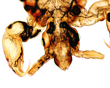
(CLICK ON IMAGES TO ENLARGE)
(20x original magnification, H&E)
(40x original magnification, H&E)
(200x original magnification, H&E)
(400x original magnification, H&E)
(200x original magnification, H&E)
Diagnosis?
A parasitologist's view of the world

2 comments:
Morphology suggestive of either myiasis or larva migrans, unable to further identify.
Florida fan
Onchocera volvulus
Post a Comment