Saturday, March 24, 2012

Case of the Week 201

The following was seen in a centrifuged urine specimen from an adopted child from Malawi. She had also been recently diagnosed with hepatitis C infection.

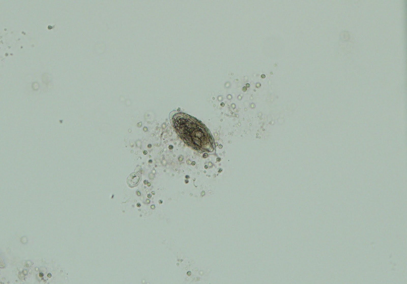
(unstained, 100 times original magnification)


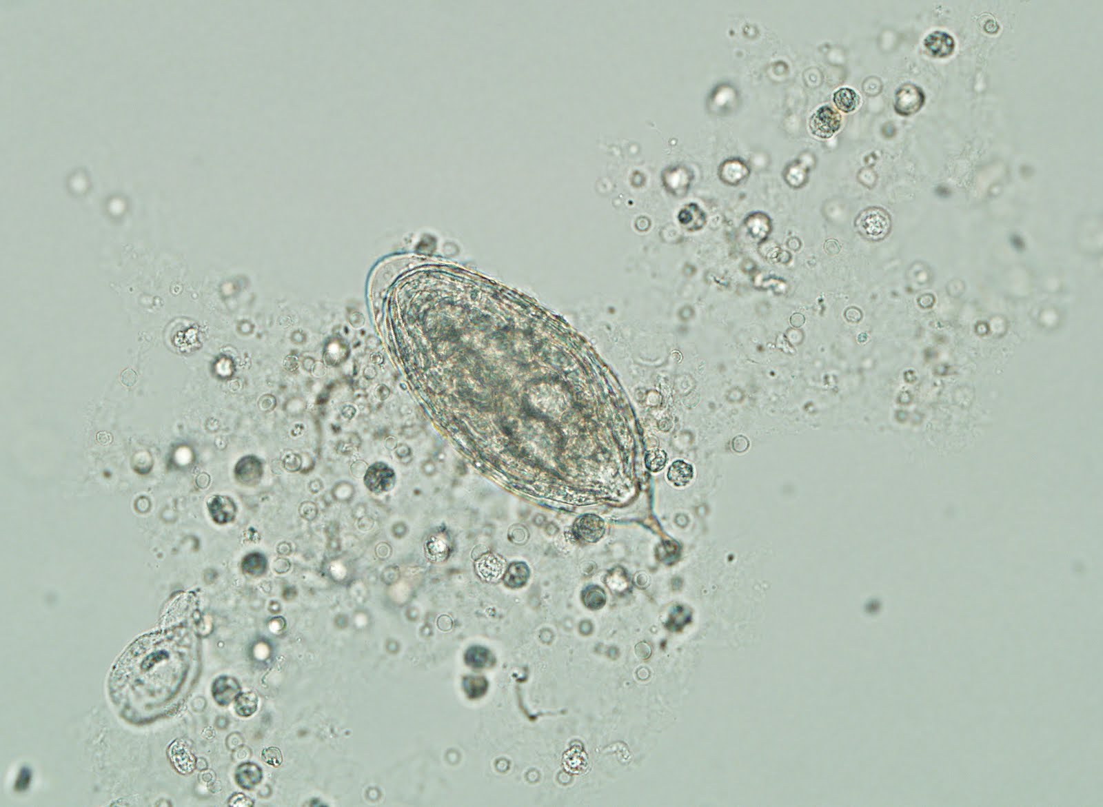
(unstained, 200 times original magnification)


(unstained, 400 times original magnification)


Identification?

This case was generously provided by MicrobeMan (Dr. Ryan Relich).

Friday, March 23, 2012

Answer to Case 201

Answer:  Schistosoma hematobium (haematobium) egg.

This egg has the characteristic terminal spine and is usually found in urine or bladder wall biopsies.  I've also seen an extensive case involving the ureters, renal pelvis of the kidneys, and the appendix (so eggs could potentially be found in stool as well).  Eggs of S. hematobium  are large, measuring 112-170 micrometers in length.

Of note, the eggs of less common species, S. intercalatum, are similar in appearance, but are larger (140-240 micrometers) in length, have a slightly longer, slightly curved terminal spine, and often have a prominent bulge in the middle of the egg (equatorial bulge). They are found in stool and not urine.

Sunday, March 18, 2012

Case of the Week 200

Welcome back to the Parasitology Case of the Week. I'm amazed to see that I am now at Case 200! I've been doing this blog for over 5 years now and have really enjoyed coming up with a weekly case and seeing your responses. Thank you for the continued support.

This week's case was generously donated by MicrobeMan.

The following object was extracted from a patient with a history of travel to a tropical rainforest over 1 month ago. He presented to a dermatologist several times since returning to the U.S. and finally the below object was extracted and sent to the parasitology lab for identification.

Identification?
Based on the organism shown, which continent would you say the patient traveled to?

Saturday, March 17, 2012

Answer to Case 200

Answer: Bot fly (human myiasis causing fly larva); specifically Dermatobia hominis

Dermatobia hominis is found in Central and South America. The larvae go through multiple instars or stages while embedded in the mammalian host. The early stages (as shown in this case) have a piriform or pear-like shape. Each larval stage has backward projecting spines encircling the thorax.

Monday, March 12, 2012

Case of the Week 199

The following arthropod was submitted for identification. The arthropod was sent to the laboratory with several information sheets from various websites including the following:
http://en.wikipedia.org/wiki/Triatominae

No history (including travel) was given but the submitting laboratory is in Wisconsin.

How would you sign this case out?

Sunday, March 11, 2012

Answer to Case 199

Identification: Leptoglossus occidentalis - Western Conifer Seed Bug (in the Family Coreidae or Leaf-footed Bugs)

Check out this BugGuide entry for more details.


My special thanks to B.A.M. from DPDx for providing the specific identification in this case.


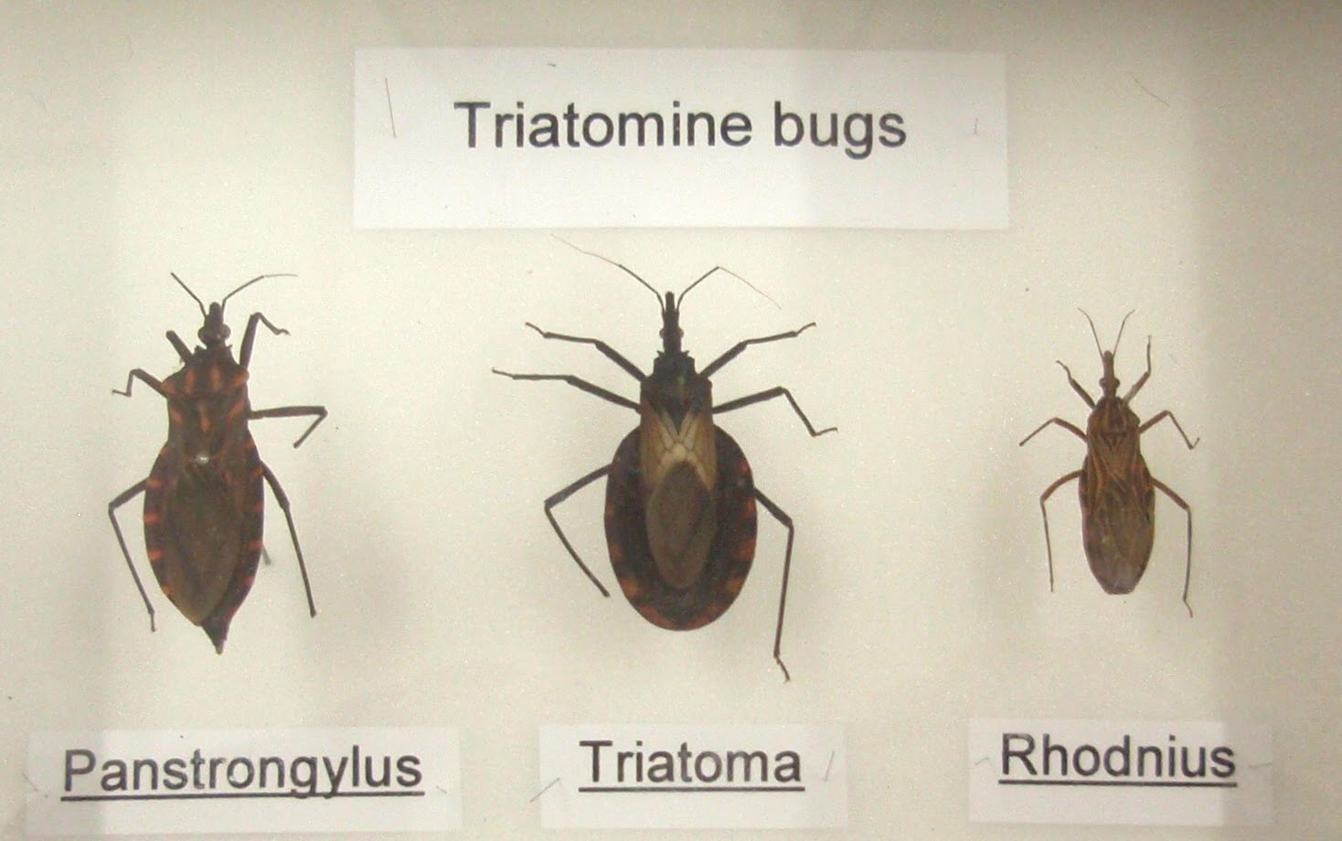
Most of you noticed that this arthropod has a clear resemblance to the "Kissing"bugs (Reduviidae family, Triatominae subfamily) that transmit the human pathogen Trypanosoma cruzi. Indeed, this is clearly what the submitting laboratory thought as well, since they included a web link to this bug along with their submission.


So herein lies the dilemma. Clinical Parasitology laboratories commonly receive both free-living and human ectoparasites for identification, and the challenge is to distinguish the two so that patients are not inappropriately treated or subjected to unnecessary eradication efforts (e.g. house fumigation, etc). Therefore, even if the clinical laboratory does not have the expertise to fully identify the non-human ectoparasites, it is important to be able to say with confidence that the object in question is NOT a human ectoparasite. The best option is to have an entomologist available for consultation. Again, DPDx is an excellent resource.


However, there are also some features that allow quick differentiation of this specimen from a kissing bug:

First, the wing venation is more numerous on this specimen (below, left) than would be seen for a kissing bug (below, right):


Second, many of the
coreids (Leaf-footed Bugs), although not all, have tibial expansions that can appear leaf-like. These can clearly be seen in this specimen and provide a quick way to exclude a kissing bug in this case.


Again, check out the BugGuide for more information.

Finally, I should point out that not all kissing bugs look alike. The subfamily Triatominae consists of 3 genera (12 species) that can transmit Trypanosoma cruzi to humans: Rhodnius, Panstrongylus, and Triatoma. These genera can be differentiated by multiple features, including placement of their antennae in relation to their eyes:

Sunday, March 4, 2012

Case of the Week 198

The following were submitted for identification by a concerned patient who had recently consumed barbequed pork ribs, along with her family. The day after, she discovered the following structures which she thought represented possible worms in the meat (CLICK ON IMAGES TO ENLARGE)

The objects were extremely hard and had blunt ends with irregular external surfaces


Also submitted was a portion of the affected pork and a histologic section:


H&E, 40 times original magnification


H&E, 100 times original magnification


H&E, 400 times original magnification


How would you sign this case out?

Saturday, March 3, 2012

Answer to Case 198

Answer: Muscle (Meat) and associated connective tissue; no parasite identified

While the long tan-white cylindrical object could potentially be mistaken for a worm, its incredibly hard nature, blunt ends and a ragged external surface is not consistent with this diagnosis and more consistent with connective tissue. The associated H&E stained section closely resembles a tendon, thus supporting the diagnosis.

Understandably, this patient was concerned with acquiring a parasite since she heard that pork potentially contained parasites. However, the more 'common' parasites found in pork worldwide are rarely encountered in the United States due to good hygienic processes and measures enforced by the USDA. These pork parasites include Taenia solium cysticerce (larvae) which are small, white-oval structures, approximately the size and shape of the head of a match, and the microscopic larvae of Trichinella spp.; neither parasite would have this gross or microscopic appearance.

Thank you everyone for the comments. They made me smile. I do indeed have a fascinating job.