Monday, December 27, 2010

Case of the Week 144

A 2 cm x 1 cm x 0.1 cm white smooth soft object was received in saline in the Parasitology lab. With manipulation (using a wooden applicator stick and wearing gloves), it was friable and fragmented easily. Some of the saline surrounding the object was pipetted onto a slide for examination, and the following objects were seen. They measure approximately 30 microns in diameter. Identification? (CLICK ON IMAGES TO ENLARGE)


Sunday, December 26, 2010

Answer to Case 144

Answer: Taenia spp. eggs. As Malassezia commented: "The hooklets are really lovely. Guessing the white smooth soft object is a proglottid?"

The white object was indeed a proglottid, despite its rather nondescript appearance and fragile nature (worm segments are usually more durable in my experience). This is why we always try to coax eggs out of possible worms that are submitted to the laboratory for identification. The presence of eggs is very useful in cases where the morphologic features of the gross object are indistinct.

In this case, these eggs have classic features of Taenia eggs, including small size (approx 30 microns diameter), thick wall with radial striations, and beautifully defined internal hooklets.

Unfortunately, it is not possible to differentiate the different Taenia spp. by their egg morphology alone, and examination of the scolex or intact proglottid is necessary. Given the fragile nature of this proglottid, further identification was not possible.

Monday, December 20, 2010

Happy Holidays!

Creepy Dreadful Wonderful Parasites wishes you a Very Happy Holidays and New Year!


Sunday, December 19, 2010

Holiday Parasite

Shown above in a Santa Claus hat is a trophozoite of Giardia intestinalis.

Monday, December 13, 2010

Case of the Week 143

The following were identified on hair clippings from a 4 year old boy. Identification?

Sunday, December 12, 2010

Answer to Case 143

Answer: Pediculus humanus egg and 1st instar nymph. The nymph had just hatched from an another egg (not shown). Note that the egg in the image still contains an unhatched larva.

Monday, December 6, 2010

Case of the Week 142

The following was seen in tissue sections of an appendix from a 10 year old boy:
(CLICK ON IMAGES TO ENLARGE). Thanks to Dr. Abdel Elhosseiny for sharing this case with us.


Diagnosis?

Sunday, December 5, 2010

Answer to Case 142

Answer: As stated very nicely by MicrobeMan, "This is...a case of enterobiasis (Enterobius vermicularis). Some diagnostic features which are nicely demonstrated in the appendix cross-sections include alae, intestines, ovaries, and the hard-to-mistake eggs (which I think look like little loaves of bread). Perhaps treatment with a benzimidazole drug or pyrantel pamoate is indicated in this case. Also, prophylactic treatment of close contacts might be warranted, since this poor fellow is a probably a nidus of infection for countless others with whom he physically interacts on a daily basis."

Note that there is a granuloma and several pinworms within the actual wall of the appendix:


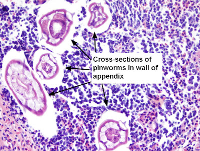
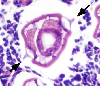
Pinworms can be identified in cross-section by their characteristic lateral alae (arrows, below), and the presence of the eggs in gravid females.


MicrobeMan also asks:
So, what's the scoop with Enterobius gregorii? Real or fiction?

Well, that's a good question. There are only scattered reports in the literature of this second Enterobius species - most over 10 years old. According to the CDC DPDx web page, "A second species, Enterobius gregorii, has been described and reported from Europe, Africa, and Asia. For all practical purposes, the morphology, life cycle, clinical presentation, and treatment of E. gregorii is identical to E. vermicularis." However, I notice that there is no other reference to this parasite on the web site. So I think for now, we will need to wait for further information to further evaluate the possibility of a second parasitic species.