Monday, December 27, 2010

Case of the Week 144

A 2 cm x 1 cm x 0.1 cm white smooth soft object was received in saline in the Parasitology lab. With manipulation (using a wooden applicator stick and wearing gloves), it was friable and fragmented easily. Some of the saline surrounding the object was pipetted onto a slide for examination, and the following objects were seen. They measure approximately 30 microns in diameter. Identification? (CLICK ON IMAGES TO ENLARGE)


Sunday, December 26, 2010

Answer to Case 144

Answer: Taenia spp. eggs. As Malassezia commented: "The hooklets are really lovely. Guessing the white smooth soft object is a proglottid?"

The white object was indeed a proglottid, despite its rather nondescript appearance and fragile nature (worm segments are usually more durable in my experience). This is why we always try to coax eggs out of possible worms that are submitted to the laboratory for identification. The presence of eggs is very useful in cases where the morphologic features of the gross object are indistinct.

In this case, these eggs have classic features of Taenia eggs, including small size (approx 30 microns diameter), thick wall with radial striations, and beautifully defined internal hooklets.

Unfortunately, it is not possible to differentiate the different Taenia spp. by their egg morphology alone, and examination of the scolex or intact proglottid is necessary. Given the fragile nature of this proglottid, further identification was not possible.

Monday, December 20, 2010

Happy Holidays!

Creepy Dreadful Wonderful Parasites wishes you a Very Happy Holidays and New Year!


Sunday, December 19, 2010

Holiday Parasite

Shown above in a Santa Claus hat is a trophozoite of Giardia intestinalis.

Monday, December 13, 2010

Case of the Week 143

The following were identified on hair clippings from a 4 year old boy. Identification?

Sunday, December 12, 2010

Answer to Case 143

Answer: Pediculus humanus egg and 1st instar nymph. The nymph had just hatched from an another egg (not shown). Note that the egg in the image still contains an unhatched larva.

Monday, December 6, 2010

Case of the Week 142

The following was seen in tissue sections of an appendix from a 10 year old boy:
(CLICK ON IMAGES TO ENLARGE). Thanks to Dr. Abdel Elhosseiny for sharing this case with us.


Diagnosis?

Sunday, December 5, 2010

Answer to Case 142

Answer: As stated very nicely by MicrobeMan, "This is...a case of enterobiasis (Enterobius vermicularis). Some diagnostic features which are nicely demonstrated in the appendix cross-sections include alae, intestines, ovaries, and the hard-to-mistake eggs (which I think look like little loaves of bread). Perhaps treatment with a benzimidazole drug or pyrantel pamoate is indicated in this case. Also, prophylactic treatment of close contacts might be warranted, since this poor fellow is a probably a nidus of infection for countless others with whom he physically interacts on a daily basis."

Note that there is a granuloma and several pinworms within the actual wall of the appendix:


Pinworms can be identified in cross-section by their characteristic lateral alae (arrows, below), and the presence of the eggs in gravid females.


MicrobeMan also asks:
So, what's the scoop with Enterobius gregorii? Real or fiction?

Well, that's a good question. There are only scattered reports in the literature of this second Enterobius species - most over 10 years old. According to the CDC DPDx web page, "A second species, Enterobius gregorii, has been described and reported from Europe, Africa, and Asia. For all practical purposes, the morphology, life cycle, clinical presentation, and treatment of E. gregorii is identical to E. vermicularis." However, I notice that there is no other reference to this parasite on the web site. So I think for now, we will need to wait for further information to further evaluate the possibility of a second parasitic species.

Monday, November 29, 2010

Case of the Week 141

The following were seen in a stool specimen (wet preparation) from a 43 year old from the Philippines. They measure approximately 70-100 µm long by 55-64 µm wide.
(CLICK ON IMAGES TO ENLARGE)

(40x magnification, unstained)


Identification?

Sunday, November 28, 2010

Answer to Case 141

Identification: Schistosoma japonicum ova

The key to making the identification is recognizing the overall shape and size (oval non-operculate eggs that are too large to be hookworm), lack of internal hooklets (as would be seen in Hymenolepis nana and H. diminuta, and finally, the presence of a small, but distinct lateral rudimentary knob, as shown here:


A rudimentary spine is not always visible on S. japonicum eggs, but the other features (size, shape, lack of hooklets) will still allow for a presumptive diagnosis.

Monday, November 22, 2010

Case of the Week 140

The following was identified in a package of frozen cod fillets purchased at a chain grocery store. (CLICK ON IMAGES TO ENLARGE)


Identification?
Does this finding pose a risk to someone who consumes this fish?

Sunday, November 21, 2010

Answer to Case 140

Answer: Anisakid worm ( including Anisakis spp. or Pseudoterranova decipiens).

To see this worm in its "cooked" state, take a peak at my previous posting HERE

These worms are nematodes in the family Anisakidae. They parasitize fish (most commonly cod, salmon, herring, mackerel, haddock, pollock, and halibut) and squid and may be transmitted to humans who eat the undercooked or raw contaminated flesh. Ingestion of the larval anisakids may cause transient human infection and possible allergic reactions in susceptible individuals. Pseudoterranova were previously known as Phocanema and Terranova (as mentioned by Anonymous).

In the infected fish, P. decipiens are brownish in color, while Anisakis spp. are red-pink. Both are typically seen in the distinctive watch-spring coil shape, and are 1.5 - 2 cm long when uncoiled. For definitive identification, examination of internal structures, including the esophagus, are necessary.

To answer the second part of the question "Does this finding pose a risk to someone who consumes this fish?":

Anisakid larvae are killed by freezing, so this worm should be non-infectious if it had been thoroughly frozen. The FDA recommends that all shellfish and fish intended for raw consumption be blast frozen to -35°C or below for fifteen hours or be frozen at -20°C or below for seven days. Anisakiasis can also be easily prevented by adequate cooking at temperatures greater than sixty degrees (until the flesh is white and flaky). It is important to note that salting and marinating will not necessarily kill the parasites.

Bon appetite!

Sunday, November 14, 2010

Case of the Week 139

The following were seen by a patient in his stool and sent to the lab for identification. How would you sign this case out? What advice should be given to the patient?
(CLICK ON IMAGES TO ENLARGE)


Saturday, November 13, 2010

Answer to Case 139

Answer: Plant material (probable derived from bananas)

Congratulations to everyone who wrote in - you all got this one right! Alasdair rightly said "Is that not just plant material? The beginnings of parasitosis of the delusional variety if it is. Advise the patient not to worry."

While these structures are commonly referred to as banana 'seeds', the Cavendish bananas that are most often sold in grocery stores do not actually develop seeds. They have been bred to be sterile. However, each aborted ovum has a vascular network consisting of xylem strands and associated cells containing astringent tannins. Upon ripening, the tannins polymerize into a semi-solid mass called 'tannin bodies' which fill the cells. The tannin bodies sometimes incorporate red-brown pigments from polyphenol oxidase activity (like the browning reaction in cut apples) as the cells age, and can therefore be seen as the red-brown bodies in this case. They are associated with the xylem strand which give them a chain-like appearance.

Anonymous Anonymous also commented "They resemble arthroconidia still stuck together. Is there a reference book for such things?"

There are a few references out there you might find helpful.

In my opinion, the best reference is the "Atlas of Human Parasitology" by Ash and Orihel which has a nice section on parasitology mimics and artifacts (although it incorrectly calls the structures in the case banana 'seeds'). These authors also have another excellent atlas called "Parasites in Human Tissues" which has a section on parasite mimics. These 2 atlases are a MUST-have for those interested in parasitology.

Another book that has some nice images is "Atlas of Microscopic Artifacts and Foreign Materials" by I-Tien Yeh.

Finally, here is a link to a short paper I wrote a while back that shows the histologic appearance of a peanut, blueberry, lentil, and hotdog: Pritt et al.

Monday, November 8, 2010

Case of the Week 138

A section of small intestine was resected from a 50 year old man from China with widespread malignancy. What infection is shown here?
(CLICK ON IMAGES TO ENLARGE)


H&E, 20x original magnification


H&E, 40x original magnification


H&E, 100x original magnification


H&E, 100x original magnification


H&E, 200x original magnification


H&E, 200x original magnification


H&E, 400x original magnification


Given the geographic origin of this individual, what is/are the most likely species involved?

Sunday, November 7, 2010

Answer to Case 138

Answer: Schistosomiasis
As Anonymous (x 2) point out, the geographic distribution is most consistent with infection due to Schistosoma japonicum. This is supported by the fact that no obvious spines are visible on any of the eggs seen. However, I should point out that seeing spines of S. mansoni or S. hematobium in tissue is often difficult due to the fact that the eggs are cut in various planes and are semi-collapsed, so that the egg outline is jagged and irregular.

Sunday, October 31, 2010

Case of the Week 137

This following amazing case was generously donated by Dr. Donald Jungkind at Thomas Jefferson University Hospital, Philadelphia, PA

The following objects were seen in a CMV shell vial viral culture from a bronchial aspiration.


(CLICK ON IMAGES TO ENLARGE)

Shell vial


Iodine-stained preparation:


Evidence of this parasite was also seen on the blood agar plate for bacterial culture.


Identification?

Saturday, October 30, 2010

Answer to Case 137

Answer: Strongyloides larvae identified in a respiratory specimen.

Although larvae in a respiratory specimen could be due to a number of different organisms, including hookworm, Ascaris, and Strongyloides, the overall length of the worm and clinical history were most consistent with Strongyloides.

The larvae may be identified by morphologic exam on stool or sputum, as well as by stool culture and concentration methods. Culture has been shown to be the most sensitive, and should be considered when there is a suspicion of strongyloidiasis. For culture, a stool or respiratory specimen containing bacteria is placed on an agar plate and incubated. A clear or blood agar can be used. The larvae will move through the agar, dragging the bacteria along with them, creating the macroscopic tracks that are visible in this case (below). Note that other worms, including hookworm, will also cause this phenomenon.


This amazing case further emphasizes the potential infectious nature of all human specimens that are submitted to the laboratory. Both the shell vial and the bacterial agar plate shown here contain infectious larvae that could penetrate intact skin and cause human infection. For this reason, it is important for all microbiologists to be familiar with the appearance of Strongyloides tracks and larvae and use appropriate infection control measures. Thanks again to Dr. Donald Jungkind for sharing this unusual case!

Sunday, October 24, 2010

Case of the Week 136

The following punch biopsy was submitted for identification of an ectoparasite.
(CLICK ON IMAGES TO ENLARGE)


Identification?
What is the preferred method for removing this ectoparasite?

Saturday, October 23, 2010

Answer to case 136

Answer: Ixodes spp. tick
As FP from VT mentioned,
"This is an Ixodes hard tick. Could be scapularis, scutal plate is the right shape. Need to see the capitulum. If this tick came thru our lab, without the capitulum we would call it Ixodes sp. unable to r/o scapularis."

Of course, I. scapularis is a tick of concern, since it is the vector of anaplasmosis, Lyme disease, and babesiosis. It is also a possible vector of the Ehrlichia muris-like organism that was recently described from Minnesota and Wisconsin.

What is the preferred method for removing this ectoparasite?
As Anonymous said:
"Best way to remove it is grab the tick by the head using forceps and pulling straight out. Being careful not to break apart."

I showed this case as an example of the way NOT to remove a tick. Forceps will do an acceptable job, and there is no need to perform a punch biopsy (which I think we would all agree is overkill)!

Monday, October 18, 2010

Case of the Week 135

The following objects were seen on a stool ova and parasite examination from a patient with diarrhea and abdominal pain. They measure approximately 80 micrometers in greatest dimension. Identification? What feature makes this parasite unique?


Sunday, October 17, 2010

Answer to Case 135

Answer: Balantidium coli trophozoites.

These protozoa are distinguished by their large size, cilia, and "kidney-bean" shaped nucleus.

What feature makes this parasite unique? They are the only ciliated protozoan to infect humans.

Sunday, October 10, 2010

Case of the Week 134

The following were an incidental finding at autopsy. Shown are hematoxylin and eosin stained sections of human tongue. (CLICK ON IMAGES TO ENLARGE)

100x original magnification


200x original magnification


200x original magnification

Diagnosis?

What is the most common source of infection worldwide? What about in the United States?

Saturday, October 9, 2010

Answer to Case 134

Answer, Part I: Trichinella spp. Although Trichinella spiralis is the most common species to infect humans in the United States, it is generally not possible to speciate based on morphologic features. The exception is Trichinella pseudospiralis whose larvae are not encapsulated compared to T. spiralis, T. nativa, T. nelsoni, T. britovi, and T. murrelli which do have encapsulated larvae. These species are the predominant to infect humans.

Answer, Part II: What is the most common source of infection worldwide? Domestic pigs. What about in the United States? Wild game

According to a publication by the Centers for Disease Control and Prevention (Roy et al."Trichinellosis Surveillance --- United States, 1997--2001." Centers for Disease Control and Prevention), trichinellosis has been steadily decreasing in the United States due to tightened regulations on pig farming and pork processing. They state:

"Although trichinellosis was associated historically with eating Trichinella-infected pork from domesticated sources, wild game meat was the most common source of infection during 1997--2001. During this 5-year period, 72 cases were reported to CDC. Of these, 31 (43%) cases were associated with eating wild game: 29 with bear meat, one with cougar meat, and one with wild boar meat. In comparison, only 12 (17%) cases were associated with eating commercial pork products, including four cases traced to a foreign source. Nine (13%) cases were associated with eating noncommercial pork from home-raised or direct-from-farm swine where U.S. commercial pork production industry standards and regulations do not apply."

The following is an excellent source of information on trichinellosis with beautiful photos: http://www.trichinella.org/index_synopsis.htm

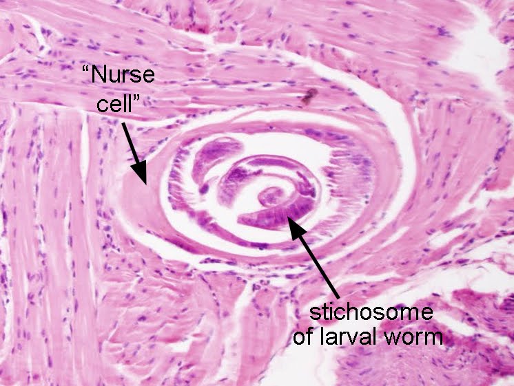
To make the diagnosis of trichinellosis on tissue section, one needs to recognize the classic appearance of the coiled larvae within tissue (typically skeletal muscle). Here is an image from this case that nicely demonstrates the larva, nurse cell (derived by the host) and stichosome (column of large rectangular cells).
(hematoxylin and eosin stained tissue section, 200x original magnification)


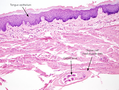
Note that in this case, the larva is located in the skeletal muscle of the tongue, right below the tongue's epithelium. (CLICK ON IMAGE TO ENLARGE)
(hematoxylin and eosin stained tissue section, 100x original magnification)


It is easiest to appreciate the coiled nature of the larvae by pressing non-fixed infected muscle between 2 slides and examining the tissue under the microscope. Here is an image from a previous case of the week that demonstrates a 'squash' prep:


Finally, for those of you non-pathologists who are having a hard time envisioning how a 3-dimensional coiled worm became a series of circles and ovals in tissue section, I've created the following diagram. The top image shows a coiled worm that is being cut along its longitudinal axis while making a slide. The bottom image shows how the worm would appear if you are only looking at the part that was cut and put on a slide.


Thanks to everyone who wrote in with the answer for this case!

Sunday, October 3, 2010

Case of the Week 133

The following was seen in an unstained wet preparation of stool from a patient complaining of abdominal pain (400X original magnification). It measures 30 microns in diameter. (CLICK ON IMAGE TO ENLARGE)


Identification?
What is a serious complication of infection with this organism?

Saturday, October 2, 2010

Answer to Case 133

Answer: Taenia spp. ovum

I also asked "What is a serious complication of infection with this organism?"

Neuro Nurse answered this very well with the following:

"Cysticercosis results from ingesting T. solium eggs. Neurocysticercosis is the most common cause of seizure disorder in South and Central America.

Passing Taenia eggs in stool is indicative of tapeworm infection from eating meat with encysted larvae. Individuals passing T. solium eggs may infect themselves and others, causing outbreaks of cysticercosis.

Eggs of Echinococcus spp. are indistinguishable from those ofTaenia spp., but humans are intermediate hosts and do not pass Echinococcus eggs in stool."

I couldn't have said it better myself! Nice Job.

I will just comment that one cannot differentiate the eggs of T. solium (which may cause cysticercosis in humans) from the eggs of T. saginata (which does NOT cause human cysticercosis). Both eggs are small (approximately 30 microns in diameter), have a classic thick wall with radial striations, and may demonstrate internal hooklets (not well appreciated in this case).

The only way to definitively distinguish the T. solium from T. saginata is by examination of the gravid proglottids or scolex, since each have characteristic morphologic features. Epidemiologic features, such as history of eating undercooked pork or beef may also be helpful to determine which parasite is more likely to cause the infection.

Monday, September 27, 2010

Case of the Week 132

The following were seen on splenic aspirate. Diagnosis?